- Biomarker-Driven Lung Cancer
- HER2 Breast Cancer
- Chronic Lymphocytic Leukemia
- Small Cell Lung Cancer
- Renal Cell Carcinoma

- CONFERENCES
- PUBLICATIONS
- CASE-BASED ROUNDTABLE

Case 1: 72-Year-Old Woman With Small Cell Lung Cancer

EP: 1 . Case 1: 72-Year-Old Woman With Small Cell Lung Cancer
Ep: 2 . case 1: extensive-stage small cell lung cancer background, ep: 3 . case 1: impower133 trial in small cell lung cancer, ep: 4 . case 1: caspian trial in extensive-stage small cell lung cancer, ep: 5 . case 1: biomarkers in small cell lung cancer, ep: 6 . case 1: small cell lung cancer in the era of immunotherapy.

EP: 7 . Case 2: 67-Year-Old Woman With EGFR+ Non–Small Cell Lung Cancer
Ep: 8 . case 2: biomarker testing for non–small cell lung cancer, ep: 9 . case 2: egfr-positive non–small cell lung cancer, ep: 10 . case 2: flaura study for egfr+ metastatic nsclc, ep: 11 . case 2: egfr+ nsclc combination therapies.

EP: 12 . Case 2: Treatment After Progression of EGFR+ NSCLC

EP: 13 . Case 3: 63-Year-Old Man With Unresectable Stage IIIA NSCLC
Ep: 14 . case 3: molecular testing in stage iii nsclc, ep: 15 . case 3: chemoradiation for stage iii nsclc, ep: 16 . case 3: pacific trial in unresectable stage iii nsclc, ep: 17 . case 3: standard of care in unresectable stage iii nsclc, ep: 18 . case 3: management of immune-related toxicities in stage iii nsclc.
Mark Socinski, MD: Thank you for joining us for this Targeted Oncology ™ Virtual Tumor Board ® focused on advanced lung cancer. In today’s presentations my colleagues and I will review three clinical cases. We will discuss an individualized approach to treatment for each patient, and we’ll review key clinical trial data that impact our decisions. I’m Dr. Mark Socinski from the AdventHealth cancer institute in Orlando, Florida. Today I’m joined by Dr Ed Kim, a medical oncologist from the Levine Cancer Institute in Charlotte, North Carolina; Dr Brendon Stiles, who is a thoracic surgeon from the Weill Cornell Medical Center in New York ; and Dr Tim Kruser, radiation oncologist from Northwestern Medicine Feinberg School of Medicine in Chicago. Thank you all for joining me today. We’re going to move to the first case, which is a case of small cell lung cancer. I’m going to ask Dr Kim to do the presentation.
Edward Kim, MD: Thanks, Mark. It’s my pleasure to walk us through the first case, which is small cell lung cancer. This is a case with a 72-year-old woman who presents with shortness of breath, a productive cough, chest pain, some fatigue, anorexia, a recent 18-pound weight loss, and a history of hypertension. She is a schoolteacher and has a 45-pack-a-year smoking history; she is currently a smoker. She is married, has 2 kids, and has a grandchild on the way. On physical exam she had some dullness to percussion with some decreased-breath sounds, and the chest x-ray shows a left hilar mass and a 5.4-cm left upper-lobe mass. CT scan reveals a hilar mass with a bilateral mediastinal extension. Negative for distant metastatic disease. PET scan shows activity in the left upper-lobe mass with supraclavicular nodal areas and liver lesions, and there are no metastases in the brain on MRI. The interventional radiographic test biopsy for liver reveals small cell, and her PS is 1. Right now we do have a patient who has extensive-stage small cell lung cancer. Unfortunately, it’s what we found. It’s very common to see this with liver metastases.
Transcript edited for clarity.

FDA Approval Marks Amivantamab's Milestone in EGFR+ NSCLC
In this episode, Joshua K. Sabari, MD, discusses the FDA approval of amivantamab plus chemotherapy as a first-line treatment for patients with EGFR exon 20 insertion mutation-positive non-small cell lung cancer.

FDA Grants Zongertinib Breakthrough Therapy Designation in HER2-Mutant NSCLC
New data on zongertinib for HER2-positive non–small cell lung cancer will be presented at the IASLC 2024 World Conference on Lung Cancer, shedding light on its potential as a novel treatment option for this patient population.

Lisberg Discusses Dato-DXd's Role in Advanced Lung Cancer Care
In this episode of Targeted Talks, Aaron Lisberg, MD, discusses results from the phase 3 TROPION-Lung01 study of datopotamab in advanced or metastatic non–small cell lung cancer.

Long-Term Immune Checkpoint Inhibition Shows Potential Extended Survival in NSCLC
Biagio Ricciuti, MD, discussed findings from a retrospective study exploring the use of immune checkpoint inhibitors for longer than 2 years in patients with non–small cell lung cancer.

Amivantamab/Lazertinib Shows Potential in Atypical EGFR-Mutant Lung Cancer
Byoung Chul Cho, MD, PhD, discussed findings from cohort C of the CHYRSALIS-2 study exploring amivantamab plus lazertinib in patients with non–small cell lung cancer with uncommon EGFR mutations.
2 Commerce Drive Cranbury, NJ 08512
609-716-7777

- Case report
- Open access
- Published: 19 August 2022
Triple primary lung cancer: a case report
- Hye Sook Choi ORCID: orcid.org/0000-0001-8387-4907 1 &
- Ji-Youn Sung 2
BMC Pulmonary Medicine volume 22 , Article number: 318 ( 2022 ) Cite this article
3077 Accesses
4 Citations
Metrics details
The risk of developing lung cancer is increased in smokers, patients with chronic obstructive pulmonary disease, individuals exposed to environmental carcinogens, and those with a history of lung cancer. Automobile exhaust fumes containing carcinogens are a risk factor for lung cancer. However, we go through life unaware of the fact that automobile exhaust is the cause of cancer. Especially, in lung cancer patient, it is important to search out pre-existing risk factors and advice to avoid them, and monitor carefully for recurrence after treatment.
Case presentation
This is the first report of a case with triple lung cancers with different histologic types at different sites, observed in a 76-year-old parking attendant. The first adenocarcinoma and the second squamous cell carcinoma were treated with stereotactic radiosurgery because the patient did not want to undergo surgery. Although the patient stopped intermittent smoking after the diagnosis, he continued working as a parking attendant in the parking lot. After 29 months from the first treatment, the patient developed a third new small cell lung cancer; he was being treated with chemoradiation.
Conclusions
New mass after treatment of lung cancer might be a multiple primary lung cancer rather than metastasis. Thus, precision evaluation is important. This paper highlights the risk factors for lung cancer that are easily overlooked but should not be dismissed, and the necessity of discussion with patients for the surveillance after lung cancer treatment. We should look over carefully the environmental carcinogens already exposed, and counsel to avoid pre-existing lung cancer risk factors at work or residence in patients with lung cancer.
Peer Review reports
The risk factors for lung cancer include smoking and inhaling exhaust fumes. Primary lung cancer (PLC) increases the risk of secondary lung cancers by four to six times [ 1 , 2 ]. With increasing exposure to environmental risk factors such as automobile exhaust fumes and advances in computed tomographic (CT) screening and treatment modality of lung cancer, the incidence of multiple primary lung cancers (MPLC) is increasing [ 2 ]. Synchronous MPLC is defined as a new cancer if it occurs with the same histology within 2 years after the PLC therapy, or with a different histology at the same time [ 3 ]; Metachronous MPLC is defined as a new cancer with the same histology if it occurs after a tumor-free period of 2 years; otherwise, it is considered to have a different histology [ 3 ]. Incidence of MPLC is higher in women, people with history of malignant disease, and those with chronic obstructive pulmonary disease (COPD), compared to solitary PLC. Men, smokers, patients with COPD, and those with non-adenocarcinomas have higher incidence of metachronous MPLC. Female sex and not smoking are independent risk factors for synchronous MPLC [ 4 ]. It is important to manage the risk factors for MPLC in patients diagnosed with lung cancer. However, patients counselling to avoid the already existing risk factors for lung cancers is not generally conducted in depth. For the first time, we report a case of triple lung cancers with metachronous MPLC in a parking attendant.
A 76-year-old man was referred for a lung mass in December 2018. He was a smoker (30 pack years with intermittent stops) and parking attendant for 30 years. There was no history of lung cancer in the immediate family of the patient. The patient was administered a dual bronchodilator for COPD.
CT scan showed a 1.4 cm × 1.3 cm mass in the right upper lobe (RUL) (Fig. 1 a) and a right lower lobe (RLL) mass-like consolidation (Fig. 1 b). Histopathologic examinations of CT-guided-percutaneous needle biopsy (PCNB) of the RUL mass revealed adenocarcinoma (ADC) (Fig. 2 a–c) with clinical staging cT1bN0M0 on ultrasonic-guided transbronchial needle biopsy (EBUS-TBNB) and fluorodeoxyglucose F18-positron emission tomography (FDG-PET) scan. RLL mass showed no metabolism on the FDG-PET scan. The FEV 1 was 56% of the predicted value. We planned a lobectomy for the RUL cancer and a follow-up for the RLL mass. However, the patient refused to undergo surgery and was treated with stereotactic radiosurgery (SRS) on the RUL mass in January 2019. The RLL mass-like consolidation did not show any changes on the follow-up chest CT or FDG-PET scan in November 2019.
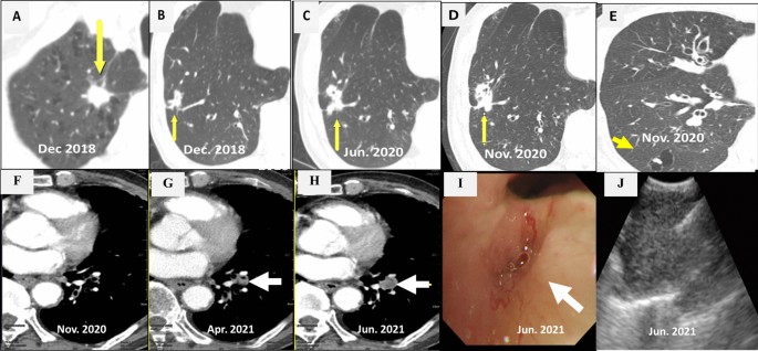
Chest CT scans. a A mass on the RUL of the first adenocarcinoma (arrow). b A mass on the RLL at the same time of the first cancer diagnosis (arrow). c Increased RLL mass six months later (arrow). d Further increased RLL mass after five months (arrow). e New nodule on the peripheral RLL (arrow). f–h Development and increase of the lymph node (arrow). i Bronchoscopic finding showing LLL anterobasal segment obstruction (arrow). j Lymph node enlargement on the EBUS. CT, computed tomography; RUL, right upper lobe; RLL, right lower lobe; LLL, left lower lobe; EBUS, endobronchial ultrasound
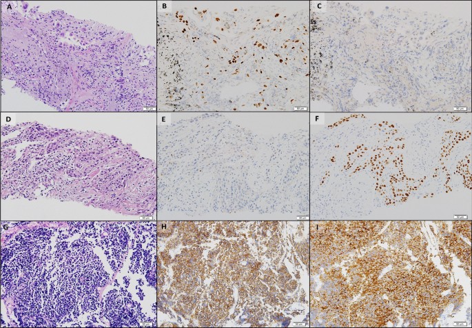
Histopathologic comparisons of the triple lung cancers. a-c The first tumor of adenocarcinoma at the right upper lobe. a Pleomorphic neoplastic cells with an acinar pattern (hematoxylin and eosin stain, ×200). b Immunoreactivity for TTF-1(×200). c Negative for P40(×200). d-f The second tumor of squamous cell carcinoma at the right lower lobe. d Polygonal cells with a solid pattern and no keratinization (hematoxylin and eosin stain, ×200). e No immunoreactivity for TTF-1(×200). f Strong staining of P40 at tumor cells(×200). g-i The third tumor of small cell carcinoma at the left lower lobe. g Small cells with scant cytoplasm and lack of nucleoli with a high mitotic activity (hematoxylin and eosin stain, ×200). h Positive neuroendocrine markers of CD56(×200). i Positive neuroendocrine marker of synaptophysin(×200). Equipment used to obtain images: Olympus BX53 microscope/Olympus objective lens WHN10X/22 UIS2, Olympus DP72 cameras and acquisition software: Olympus CellSens Standard 1.6 software. TTF-1, thyroid transcription factor-1
In June 2020, the RLL mass-like consolidation was found to have increased on a chest CT scan (Fig. 1 c). PCNB of the RLL mass was performed, and histologic examination revealed anthracofibrosis. Five months later, the RLL mass increased further (Fig. 1 d), and a new nodule appeared at the periphery of the RLL (Fig. 1 e). PCNB was performed again on the same RLL mass (Fig. 1 d), and histological examination demonstrated squamous cell carcinoma (SCC) (Fig. 2 d–f). There was no metastasis except for hypermetabolism of the new nodule in the RLL periphery (Fig. 1 e) on the FDG-PET scans. We could not perform a biopsy for the new peripheral nodule (Fig. 1 e) due to cystic changes. We concluded the clinical staging of the RLL SCC as cT3N0M0 on the EBUS-TBNB and PET scan. SRSs were performed separately for the RLL SCC and the new RLL peripheral nodule, respectively in February 2021.
We performed chest CT scan for surveillance of lung cancer. Five months later after 2nd SCC diagnosis, a new nodule emerged at the left lower lobe (LLL) (Fig. 1 f, g). Two months after that, the nodule increased further (Fig. 1 h). Bronchoscopy showed new total obstruction of the anterobasal segmental bronchus of the LLL (Fig. 1 i). Histologic examinations of bronchial biopsy and EBUS-TBNB (Fig. 1 j) for LLL lesions demonstrated small cell lung carcinoma (SCLC) (Fig. 2 g–i). Clinical staging was limited stage. The patient was treated with chemotherapy (etoposide/carboplatin) and concurrent thoracic radiation.
Discussion and conclusions
Smoking is a notorious risk factor for lung cancer. The parking attendant was exposed to exhaust fumes, including carcinogens from the fuel. He was using a bronchodilator for COPD. Smoking and COPD are independent risk factors for MPLC [ 4 ]. PLC increased the risk of MPLC despite stage IA lung cancer [ 5 , 6 ]. We suggest that his history of exposure to exhaust fumes in addition to smoking, COPD, and PLC contributed to the metachronous MPLC.
At the time of the first ADC diagnosis on the RUL, we discuss the possibility that the RLL mass was lung cancer, and decided to follow according to the PET-CT scan results with the multidisciplinary approach. Unfortunately, 18 months later, PCNB and histologic findings for the RLL mass showed no cancers. Five months after that (23 months after the first ADC treatment), repeated PCNB on the RLL mass demonstrated SCC. The possibility that an additional abnormality is cancer must be addressed when PLC is diagnosed.
The third SCLC of LLL developed newly 29 months after the first ADC treatment. It was detected after 5 months after the diagnosis of second cancer. Timely CT scan for surveillance is essential for earlier diagnosis of metachronous MPLC in the patients with PLC, which could be improve the outcomes of MPLC. We considered that the first ADC and the second SCC were synchronous MPLC; thus, the third SCLC might be metachronous MPCL. The three different types of MPLC were not a transformation of the PLC after SRSs, but originally developed from three different histologies. Recently, genetic/molecular profiles have begun to be used for differentiation and diagnosis of MPLC [ 7 ]. and further investigation is needed.
The primary tumor control rate of SRS is 97.6% in medically inoperable early-stage non-SCLC [ 8 ]. Recently, the risk of metachronous MPLC was found to be lower with radiotherapy than non-radiotherapy [ 6 , 8 ] even though in stage IA lung cancer [ 5 ]. The incidence of metachronous MPLC was 0.5% at 1 year and 2.28% at 5 years among solitary PLC survivors with radiotherapy, which was lower compared to the non-radiotherapy group [ 6 ]. Based on these findings, it is assumed that the SRSs might not induce metachronous MPLC in our patient.
The question was what could have been responsible for the patient’s triple lung cancers. Unknown susceptible genetic factors, smoking, and exhaust fumes might have contributed to the development of triple lung cancers. Previously reported risk factors [ 4 ] such as male sex, smoking, COPD, and nonadenocarcinoma also increased the risk of metachronous MPLC in this patient. He stopped smoking after the first diagnosis of lung cancer, but continued as a parking attendant for 12 h a day. It is well known that harmful effects of smoking persist for years even after smoking cessation. Thus, the main cause of lung cancer in this patient is likely to be smoking. Physicians always counsel their lung cancer patients that smoking is one of the main causes of lung cancer and advise to quit smoking immediately. However, the emphasis on counselling avoidance of other environmental carcinogens that may have a synergistic effect with smoking is often neglected. This patient was exposed to exhaust gas at work for 30 years which is a known occupational carcinogen, and exposure continued even after quitting smoking and diagnosing lung cancer. He had no family history of lung cancer. Unfortunately, his wife was diagnosed with stage IV lung adenocarcinoma lung cancer at August 2021, the time of 3 rd SCLC diagnosis of him. He and his wife had worked together in parking lot for several years. We suggest that exhaust fumes might be an additional main risk factor for metachronous MPLC that is easily overlooked in this patient.
Despite stage I lung cancer, careful surveillance for metachronous MPLC is needed, especially in patients with a history of smoking, COPD, PLC, and exposure to environmental carcinogens such as exhaust fumes. Occupation and environment surveys with attentive advice for risk factors of lung cancer are very important, and it is valuable to evaluate concurrent abnormal images in patients with lung cancer. Appropriate CT scan surveillance after PLC therapy can help identify curable MPLC, which might lead to improved overall survival.
Availability of data and materials
All data generated or analyzed during this study are included in this published article.
Abbreviations
Adenocarcinoma
Chronic obstructive pulmonary disease
Computed tomography
Ultrasonic-guided transbronchial needle biopsy
F18-positron emission tomography
Left lower lobe
Primary lung cancer
Multiple primary lung cancers
Percutaneous needle biopsy
Right lower lobe
Right upper lobe
Squamous cell carcinoma
Small cell lung carcinoma
Stereotactic radiosurgery
Johnson BE. Second lung cancers in patients after treatment for an initial lung cancer. J Natl Cancer Inst. 1998;90(18):1335–45.
Article CAS Google Scholar
Surapaneni R, Singh P, Rajagopalan K, Hageboutros A. Stage I lung cancer survivorship: risk of second malignancies and need for individualized care plan. J Thorac Oncol. 2012;7(8):1252–6.
Article Google Scholar
Martini N, Melamed MR. Multiple primary lung cancers. J Thorac Cardiovasc Surg. 1975;70(4):606–12.
Shintani Y, Okami J, Ito H, Ohtsuka T, Toyooka S, Mori T, Watanabe S-i, Asamura H, Chida M, Date H, et al. Clinical features and outcomes of patients with stage I multiple primary lung cancers. Cancer Sci. 2021;112(5):1924–35.
Khanal A, Lashari BH, Kruthiventi S, Arjyal L, Bista A, Rimal P, Uprety D. The risk of second primary malignancy in patients with stage Ia non-small cell lung cancer: a US population-based study. Acta Oncol. 2018;57(2):239–43.
Hu ZG, Tian YF, Li WX, Zeng FJ. Radiotherapy was associated with the lower incidence of metachronous second primary lung cancer. Sci Rep. 2019;9(1):19283–19283.
Asamura H. Multiple primary cancers or multiple metastases, that is the question. J Thorac Oncol. 2010;5(7):930–1.
Timmerman R, Paulus R, Galvin J, Michalski J, Straube W, Bradley J, Fakiris A, Bezjak A, Videtic G, Johnstone D, et al. Stereotactic body radiation therapy for inoperable early stage lung cancer. JAMA. 2010;303(11):1070–6.
Download references
Acknowledgements
Not applicable
No funding sources were used.
Author information
Authors and affiliations.
Department of Internal Medicine, Kyung Hee Unversity Medical Center, 23 Kyunghee dae-ro, Dongdaemun-gu, Seoul, 02447, Republic of Korea
Hye Sook Choi
Department of Pathology, Kyung Hee University Medical Center, Seoul, Republic of Korea
Ji-Youn Sung
You can also search for this author in PubMed Google Scholar
Contributions
HSC drafted the manuscript, reviewed the literature, and collected the data. JYS collected the data and revised the manuscript. All authors contributed to obtaining and interpreting the clinical information. All authors read and approved the final manuscript.
Corresponding author
Correspondence to Hye Sook Choi .
Ethics declarations
Ethics approval and consent to participate.
This study was approved by the Kyung Hee University Medical Center (approval number: KHUH 2021–09-069–002) and written informed consent was given by the patient.
Consent for publication
Written informed consent was obtained from the patient for publication of this case report and any accompanying images. A copy of the written consent is available for review by the Editor-in-Chief of this journal.
Competing interests
The authors declare that they do not have any conflict of interest.
Additional information
Publisher's note.
Springer Nature remains neutral with regard to jurisdictional claims in published maps and institutional affiliations.
Rights and permissions
Open Access This article is licensed under a Creative Commons Attribution 4.0 International License, which permits use, sharing, adaptation, distribution and reproduction in any medium or format, as long as you give appropriate credit to the original author(s) and the source, provide a link to the Creative Commons licence, and indicate if changes were made. The images or other third party material in this article are included in the article's Creative Commons licence, unless indicated otherwise in a credit line to the material. If material is not included in the article's Creative Commons licence and your intended use is not permitted by statutory regulation or exceeds the permitted use, you will need to obtain permission directly from the copyright holder. To view a copy of this licence, visit http://creativecommons.org/licenses/by/4.0/ . The Creative Commons Public Domain Dedication waiver ( http://creativecommons.org/publicdomain/zero/1.0/ ) applies to the data made available in this article, unless otherwise stated in a credit line to the data.
Reprints and permissions
About this article
Cite this article.
Choi, H.S., Sung, JY. Triple primary lung cancer: a case report. BMC Pulm Med 22 , 318 (2022). https://doi.org/10.1186/s12890-022-02111-x
Download citation
Received : 07 April 2022
Accepted : 10 August 2022
Published : 19 August 2022
DOI : https://doi.org/10.1186/s12890-022-02111-x
Share this article
Anyone you share the following link with will be able to read this content:
Sorry, a shareable link is not currently available for this article.
Provided by the Springer Nature SharedIt content-sharing initiative
- Multiple primary lung cancer (MLPC)
- Synchronous MLPC
- Metachronous MLPC
- Parking attendant
BMC Pulmonary Medicine
ISSN: 1471-2466
- Submission enquiries: [email protected]
- General enquiries: [email protected]
- Get new issue alerts Get alerts
Secondary Logo
Journal logo.
Colleague's E-mail is Invalid
Your message has been successfully sent to your colleague.
Save my selection
A case report of metastatic lung adenocarcinoma with long-term survival for over 11 years
Editor(s): NA.,
Department of Respiratory Medicine, Tokyo Dental College, Ichikawa General Hospital, 5-11-13, Sugano, Ichikawa, Chiba, Japan.
∗Correspondence: Tatsu Matsuzaki, Department of Respiratory Medicine, Tokyo Dental College, Ichikawa General Hospital, 5-11-13, Sugano, Ichikawa, Chiba 272-8513, Japan (e-mail: [email protected] ).
Abbreviations: CBDCA = carboplatin, CDDP = cisplatin, CEA = carcinoembryonic antigen, DTX = docetaxel, ECOG-PS = Eastern Cooperative Oncology Group performance status, EGFR = epidermal growth factor receptor, EGFR-TKI = epidermal growth factor receptor tyrosine kinase inhibitor, NSCLC = non-small cell lung cancer, OS = overall survival, PD = progressive disease, PEM = pemetrexed, PFS = progression-free survival, RC = re-challenge chemotherapy, RR = response rate, UICC = Union of International Cancer Control.
This research did not receive any specific grant from funding agencies in the public, commercial, or not-for-profit sectors.
The Ethics Committee of our hospital approved the study and provided permission to publish the results.
The patient provided written informed consent and has provided consent for publication of the case.
The authors declare that they have no conflict of interest.
This is an open access article distributed under the Creative Commons Attribution License 4.0 (CCBY), which permits unrestricted use, distribution, and reproduction in any medium, provided the original work is properly cited. http://creativecommons.org/licenses/by/4.0
Rationale:
This is the first known report in the English literature to describe a case of metastatic non-small cell lung cancer that has been controlled for >11 years.
Patient concerns:
A 71-year-old man visited our hospital because of dry cough.
Diagnosis:
Chest computed tomography revealed a tumor on the left lower lobe with pleural effusion, and thoracic puncture cytology indicated lung adenocarcinoma.
Interventions:
Four cycles of carboplatin and docetaxel chemotherapy reduced the size of the tumor; however, it increased in size after 8 months, and re-challenge chemotherapy (RC) with the same drugs was performed. Repeated RC controlled disease activity for 6 years. After the patient failed to respond to RC, erlotinib was administered for 3 years while repeating a treatment holiday to reduce side effects. The disease progressed, and epidermal growth factor receptor ( EGFR ) gene mutation analysis of cells from the pleural effusion detected the T790 M mutation. Therefore, osimertinib was administered, which has been effective for >1 year.
Outcomes:
The patient has survived for >11 years since the diagnosis of lung cancer.
Lessons:
Long-term survival may be implemented by actively repeating cytotoxic chemotherapy and EGFR-tyrosine kinase inhibitor administration.
1 Introduction
The prognosis of patients with advanced non-small cell lung cancer (NSCLC) is poor, and their 1-year survival rate after cytotoxic chemotherapy is only 29%. [1] However, the development of epidermal growth factor receptor tyrosine kinase inhibitors (EGFR-TKIs) dramatically improves the prognosis of certain patients. Patients with EGFR-mutant advanced NSCLC receiving EGFR-TKIs have a median overall survival (OS) more than twice as long as those not receiving EGFR-TKIs (24.3 vs 10.8 months). [2] The 5-year survival rate of patients with EGFR-mutant metastatic lung adenocarcinoma treated with EGFR-TKIs is 14.6%. [3] However, metastatic NSCLC patients with long-term survival (>10 years) are still rare.
We treated an advanced NSCLC patient with malignant pleural effusion who survived for >11 years and for whom disease progression was controlled using drugs alone without surgery or radiation therapy.
2 Case presentation
A 71-year-old Japanese man experienced dry cough for 2 weeks and visited the Department of Respiratory Medicine at our hospital in August 2007. Enhanced chest-abdomen computed tomography revealed a tumor with a 3-cm diameter in the left lower lobe and left pleural effusion ( Fig. 1 ). A 5-mm nodule, considered to be lung metastasis, was detected in the left upper lobe. Cytological analysis of the left pleural effusion by thoracic puncture led to the diagnosis of lung adenocarcinoma. Gadolinium-enhanced brain magnetic resonance imaging and bone scintigraphy did not reveal any other metastases. The tumor was classified as clinical T4N0M1, stage IV according to the TNM classification of the Union of International Cancer Control (UICC), 6th edition. According to the UICC 8th edition, it was classified as clinical T4N0M1a, stage IV A. The patient had a history of hypertension and was a past smoker (60 pack-years) and a company employee. The Eastern Cooperative Oncology Group performance status (ECOG-PS) at the time of admission was 1. The carcinoembryonic antigen (CEA) level was 97.4 ng/mL (normal, 0–5 ng/ml).

Beginning in August 2007, the patient received carboplatin (CBDCA) and docetaxel (DTX). After 4 cycles, the tumor was reduced to 1 cm in diameter. The 5-mm nodule and pleural effusion had also decreased. According to the Response Evaluation Criteria in Solid Tumors version 1.1, partial response was achieved, but he experienced progressive disease (PD) after 8 months. Six cycles of re-challenge chemotherapy (RC) using the same regimen were started in August 2008 and were effective. Thereafter, at each recurrence of PD, 4 to 6 cycles of RC were administered, and by 2013, 38 cycles had been completed over 6 years of treatment ( Fig. 2 A). However, we could no longer control disease activity using the same chemotherapy regimen. Moreover, primary tumor size evaluation became difficult owing to massive pleural effusion; although not standard, we estimated the effect of treatment using the increase and decrease of CEA as an index. CEA increased from a minimum of 4.6 ng/ml to 33.3 ng/ml in October 2013 during repeated cytotoxic chemotherapy. Although his EGFR mutation status was unknown, we initiated erlotinib administration and the CEA level decreased. After 8 weeks, the patient developed grade 3 acneiform rash, assessed using the Common Terminology Criteria for Adverse Events version 5.0, and erlotinib administration was discontinued for 6 weeks. Cycles of medication and treatment holiday were repeated, and the patient was carefully observed for skin rash. Dose reduction was attempted once, but it was not effective, because we noted an elevated CEA level and intolerable skin rash. For 3 years, 4-week erlotinib administration was repeated with 4–6-week treatment holiday intervals ( Fig. 2 B). CEA increased from a minimum of 3.1 ng/ml to 30.4 ng/ml in January 2017 during treatment with erlotinib. We performed EGFR mutation analysis using adenocarcinoma cells from the pleural effusion and detected exon 19 deletion and exon 20 T790 M mutation; therefore, osimertinib was substituted for erlotinib.

We continued monthly CEA measurements after beginning osimertinib administration and noted that the level continued to decrease. In August 2018, the CEA level was 12.1 ng/ml and the ECOG-PS was 1. As of the last follow-up, the patient has survived for >11 years since the diagnosis of lung cancer.
3 Discussion
The clinical data of 10 patients with advanced NSCLC who survived for >5 years were retrospectively reviewed, and a good PS, adenocarcinoma, and a history of EGFR-TKI administration were the factors contributing to long-term survival. [4] According to another retrospective study, [3] 20 of 137 patients with EGFR-mutant lung adenocarcinoma survived for ≥5 years, and exon 19 deletion, absence of extrathoracic metastases, absence of brain metastasis, and current non-smoking status were reportedly good prognostic factors. Our case corroborated the good prognostic factors reported in these studies.
A case of metastatic NSCLC in which the patient survived for 10 years has already been reported; however, the patient underwent not only chemotherapy but also surgery and radiation therapy. [5] To the best of our knowledge, ours is the first report in the English literature to describe a metastatic NSCLC case controlled for >11 years. Moreover, our patient was only treated with chemotherapy and EGFR-TKIs.
We considered that 4 treatment policies may be the key to success:
- 1. RC with CBDCA plus DTX;
- 2. repeated re-challenge erlotinib administration;
- 3. osimertinib administration after T790 M mutation in exon 20, which confers resistance to erlotinib; and
- 4. use of both cytotoxic drugs and EGFR-TKIs. We will particularly focus on the first and second policies because of the non-standard methods.
The response rate (RR) to RC of platinum doublets containing pemetrexed (PEM) or taxanes is reportedly 27.5%, with a progression-free survival (PFS) of 3.9 months and an OS of 8.7 months. This RR is high, but the PFS and OS are similar to those seen with administration of a single-agent as second-line treatment. [6] Advanced NSCLC patients for whom RC with 2-drug combination therapy is performed have a longer median survival than those administered only DTX as second-line treatment. [7] The current evidence that RC is superior to a single agent second-line treatment is not sufficient, but if the side effects are acceptable, RC may be a suitable option.
To safely perform RC using platinum-based 2-drug therapy, it may be necessary to include a treatment holiday period for a certain duration to facilitate physical fitness recovery. Time to progression of >3 months after ending first-line chemotherapy is a predictor of long-term survival (>2 years) in advanced NSCLC patients who receive cytotoxic chemotherapy. [8] Advanced NSCLC patients who survive for >2 years have a good response to first-line cytotoxic chemotherapy, and a prolonged treatment-free interval increases long-term survival. [9] In the present case, the treatment holiday after cytotoxic chemotherapy was approximately 6 months. A prolonged treatment-free interval appears to be important for restoring physical fitness; therefore, the patient could tolerate the next treatment.
A meta-analysis of randomized control studies compared cisplatin (CDDP) and CBDCA in advanced NSCLC patients. Although regimens containing CDDP did not prolong OS, subgroup analysis demonstrated that, when combined with third-generation anticancer drugs, CDDP prolonged OS more than CBDCA in patients with non-squamous cell carcinoma. [10] In the TAX 326 trial, CBDCA and DTX combination therapy helped achieve an OS equivalent to that associated with CDDP and vinorelbine combination therapy; in addition, the CBDCA and DTX combination was well tolerated and facilitated a high quality of life. [11] Here, 38 cycles of CBDCA and DTX combination therapy were administered. To perform platinum-based 2-drug RC, it may be advantageous to repeatedly administer CBDCA rather than CDDP to facilitate tolerability.
When erlotinib toxicity is intolerable, as in our patient who developed a severe skin rash, the dosage is generally reduced. In patients with EGFR-mutant NSCLC, dose reduction (25 mg/day) of erlotinib can reduce the toxicity while maintaining efficacy. [12] In contrast, a prospective phase II trial involving low-dose erlotinib in patients with EGFR-mutant NSCLC revealed that dose reduction (50 mg/day) is not recommended because of reduced efficacy. [13] Intermittent erlotinib administration on alternate days successfully maintained efficacy while reducing toxicity. [14] Here, disease control was possible by RC of platinum doublets for approximately 6 cycles after the 6-month treatment holiday. Therefore, we applied the RC strategy to EGFR-TKI administration. Continued erlotinib administration was discontinued if side effects became intolerable or health-threatening and resumed when side effects disappeared. Toxicity and efficacy were balanced by allowing an approximately 4–6-week treatment holiday after the 4-week erlotinib administration. To the best of our knowledge, we were the first to employ erlotinib in RC; this method was named repeated re-challenge administration of erlotinib and was considered to alleviate suffering due to toxicity. Although there is little evidence to determine whether dose reduction, intermittent administration, or repeated re-challenge administration is better, it is important to avoid complete cessation of therapy.
The exon 20 mutation in EGFR , leading to the T790 M mutation, is a resistance mechanism to traditional EGFR-TKIs. Osimertinib treatment is associated with a longer median PFS than chemotherapy using PEM plus either CBDCA or CDDP as second-line treatment in T790M-mutant patients who received EGFR-TKIs. [15] Our patient with the exon 19 deletion and T790 M mutation received osimertinib after erlotinib. Even if the tumor develops resistance to conventional EGFR-TKIs, it is important to recognize that drugs such as osimertinib are available and may improve long-term survival.
In EGFR-mutant advanced NSCLC with exon 19 deletion and exon 21 (L858R) mutation, patients who receive sequential therapy with erlotinib and cytotoxic drugs experience a longer median OS than those who receive cytotoxic drugs or erlotinib alone. [16] It may be important to administer both EGFR-TKIs and cytotoxic drugs throughout the course of treatment.
Although more study is required, RC of CBDCA plus DTX and repeated re-challenge administration of erlotinib may be an empirical option for long-term survival. To understand the clinical and biological background of long-term survival cases, accumulation of future cases similar to ours is warranted.
Acknowledgments
We would like to thank Editage ( www.editage.jp ) for English language editing.
Author contributions
Supervision: Takeshi Terashima.
Writing – original draft: Tatsu Matsuzaki.
Writing – review & editing: Eri Iwami, Kotaro Sasahara, Aoi Kuroda, Takahiro Nakajima.
- Cited Here |
- Google Scholar
- View Full Text | PubMed | CrossRef |
- PubMed | CrossRef |
adenocarcinoma; EFGR-TKIs; EGFR gene mutation; long-term survival; non-small cell lung cancer; re-challenge chemotherapy; T790 M
- + Favorites
- View in Gallery
Readers Of this Article Also Read
Global and regional prevalence and burden for premenstrual syndrome and..., severe re-expansion pulmonary edema after chest tube insertion for the..., is intraoperative corticosteroid a good choice for postoperative pain relief in ..., the incidence and prognosis of thymic squamous cell carcinoma: a surveillance,..., klebsiella pneumoniae</em> k2-st86: case report', 'yamamoto hiroyuki md phd; iijima, anna ms; kawamura, kumiko phd; matsuzawa, yasuo md, phd; suzuki, masahiro phd; arakawa, yoshichika md, phd', 'medicine', 'may 22, 2020', '99', '21' , 'p e20360');" onmouseout="javascript:tooltip_mouseout()" class="ejp-uc__article-title-link">fatal fulminant community-acquired pneumonia caused by hypervirulent klebsiella ....
Log in using your username and password
- Search More Search for this keyword Advanced search
- Thorax Education
- Latest content
- Current issue
- BMJ Journals
You are here
- Volume 64, Issue 7
- A case of small cell lung cancer treated with chemoradiotherapy followed by photodynamic therapy
- Article Text
- Article info
- Citation Tools
- Rapid Responses
- Article metrics
- Department of Internal Medicine; College of Medicine, Chungnam National University Hospital and Cancer Research Institute, Jungku, Daejeon, South Korea
- Professor J O Kim, Department of Internal Medicine, Chungnam National University Hospital and Cancer Research Institute, 640 Daesadong, Jungku, Daejeon 301-721, South Korea; jokim{at}cnu.ac.kr
Here, we present the case of a 51-year-old man with limited-stage small cell lung cancer (LS-SCLC) who received concurrent chemoradiotherapy and photodynamic therapy (PDT). The patient was diagnosed as having LS-SCLC with an endobronchial mass in the left main bronchus. Following concurrent chemoradiotherapy, a mass remaining in the left lingular division was treated with PDT. Clinical and histological data indicate that the patient has remained in complete response for 2 years without further treatment. This patient represents a rare case of complete response in LS-SCLC treated with PDT.
https://doi.org/10.1136/thx.2008.112912
Statistics from Altmetric.com
Request permissions.
If you wish to reuse any or all of this article please use the link below which will take you to the Copyright Clearance Center’s RightsLink service. You will be able to get a quick price and instant permission to reuse the content in many different ways.
Competing interests: None.
Patient consent: Obtained.
Read the full text or download the PDF:

Call: 1.631.629.4328 Mon-Fri 10 am - 2 pm EST

A 75-Year-Old Female Smoker with Advanced Small-Cell Lung Cancer and Eastern Cooperative Oncology Group Performance Status 2 who Responded to Combination Immunochemotherapy with Atezolizumab, Etoposide, and Carboplatin
Unusual clinical course, Unusual setting of medical care, Educational Purpose (only if useful for a systematic review or synthesis)
- 1 Second Department of Lung Diseases and Tuberculosis, Medical University of Białystok, Białystok, Poland
- A Study design/planning
- B Data collection/entry
- C Data analysis/statistics
- D Data interpretation
- E Preparation of manuscript
- F Literature analysis/search
- * Corresponding author: [email protected]
DOI: 10.12659/AJCR.936536
Am J Case Rep 2022; 23:e936536
- * Corresponding Author: Michał Dębczyński, e-mail: [email protected]
- Submitted: 01 March 2022
- Accepted: 08 June 2022
- In Press: 01 July 2022
- Published: 11 August 2022
This paper has been published under Creative Common Attribution-NonCommercial-NoDerivatives 4.0 International ( CC BY-NC-ND 4.0 ) allowing to download articles and share them with others as long as they credit the authors and the publisher, but without permission to change them in any way or use them commercially.

BACKGROUND: Atezolizumab is an immune checkpoint inhibitor used as first-line treatment with carboplatin and etoposide chemotherapy for advanced small cell lung cancer. Immunochemotherapy treatment decisions can be affected by patients’ physical ability. Because of the exclusion of patients with an Eastern Cooperative Oncology Group Performance Status (ECOG PS) ≥2 from clinical trials, treatment outcome evidence in this group is limited.
CASE REPORT: We present the case of a 75-year-old woman with an ECOG PS of 2 admitted with respiratory symptoms and diagnosed with advanced small-cell lung cancer. After managing exacerbation of COPD and decompensated heart failure, atezolizumab with carboplatin and etoposide was administered. After 2 cycles of immunochemotherapy, deterioration of health was observed, including anemia and thrombocytopenia. Because of the good response in imaging tests and restored balance of the patient condition, immunochemotherapy was continued. After 4 cycles of combined treatment, complete regression was achieved. No another adverse effects were observed. The patient was qualified for maintenance therapy with atezolizumab. In follow-up CT scan after 2 cycles of atezolizumab, progression was observed and patient was qualified for second-line treatment.
CONCLUSIONS: This report presents the case of an older patient with advanced small cell lung cancer and an ECOG status of 2 who responded to combined immunochemotherapy with atezolizumab, etoposide, and carboplatin. Adverse effects observed during immunotherapy were not a reason for discontinuation of the therapy. The assessment of the effectiveness of immunotherapy in patients with ECOG PS ³2 is difficult owing to the insufficient representation of this group in clinical trials.
Keywords: Antineoplastic Combined Chemotherapy Protocols, case reports, Immunotherapy, Physical Functional Performance, Aged, Antibodies, Monoclonal, Humanized, Carboplatin, Etoposide, Female, Group Processes, Humans, Lung Neoplasms, smokers
Small-cell lung cancer (SCLC) accounts for 15% to 20% of all lung cancer cases. Most of the patients diagnosed with this cancer are ≥65 years old, and the median age is 67 years [1,2]. SCLC is a cancer with an unfavorable prognosis [3]. The 3-year survival rate before the combined treatment era was 12% to 25% in the limited disease stage and did not exceed 2% in the disseminated disease stage [3]. SCLC is a highly tobacco-dependent cancer, as most of the patients diagnosed with SCLC are former or current cigarette smokers [4]. It is characterized by a high mitotic index and thus a rapid growth rate. Often, distant metastases are found at diagnosis. The limited form of the disease affects only 30% of patients [5]. The main method of treating patients with SCLC is chemotherapy based on etoposide and platinum derivatives. Recently, new therapeutic options have emerged in first-line treatment, with the addition of immunological drugs to standard chemotherapy. In some cases, the addition of an immune checkpoint inhibitor drug is associated with an increase in overall survival and progression-free survival [6]. There are still ongoing medical trials being conducted to more accurately define the safety and efficacy of atezolizumab in combination with carboplatin and etoposide in patients with untreated extensive-stage SCKC [7,8]. When qualifying patients for treatment, it is very important to accurately and individually assess the degree of performance status (PS) of patients, distinguishing whether a poor PS score results from the neoplastic disease itself and its complications or from concomitant diseases [9].
PS is a tool often used by oncology healthcare professionals to assess the fitness of patients for systemic anticancer therapy and to predict prognosis in advanced malignancy. The Eastern Cooperative Oncology Group Performance Status (ECOG PS) scoring system (Table 1) is considered an effective and reliable scale, which shows no significant variations in PS assessment by different healthcare professionals [10].
In a meta-analysis, Dall’Olio et al noticed that the high level of heterogeneity for overall survival analysis in the PS ≥2 population with advanced non-small cell lung cancer (NSCLC) treated with immune checkpoint inhibitors could be the result of the patient heterogeneity within the PS 2 population and the subjectivity of the ECOG PS assessment. They indicate that poorer PS is correlated with lower immunotherapy efficacy [12].
Most case reports in the literature refer to the immunochemotherapy in NSCLC. There are reports of using chemotherapy with atezolizumab in an 80-year-old male patient with extensive-stage SCLC undergoing hemodialysis [13]. Authors of another case report also suggested that SCLC patients outside of clinical trials are typically in a poor ECOG state but could also benefit from immunotherapy [14].
In the presented study, we report a case of a 75-year-old female patient with advanced SCLC and an ECOG PS of 2 who responded well to combination immunochemotherapy with atezolizumab, etoposide, and carboplatin. We draw attention to the qualification and benefits of combination therapy: immunochemotherapy in a patient with SCLC at an older age and an ECOG PS of 2 and the impact of possible adverse effects on this type of treatment.
Case Report
A 75-year-old woman who was an active smoker (about 30 pack-years), reported reduced exercise tolerance, exercise dyspnea, and periodic chest pain. She had been experiencing a gradual deterioration in her health for about 3 weeks. The patient associated respiratory system ailments with a past infection. She was taking symptomatic medications with no significant improvement. Two weeks prior, the patient noticed hemoptysis; therefore, she went to the family doctor. She was prescribed antibiotic therapy (amoxicillin) on an outpatient basis, and experienced no noticeable improvement. Then, the patient was referred to the Department of Lung Diseases.
Reported comorbidities included chronic obstructive pulmonary disease, arterial hypertension, ischemic heart disease, and previous infection with SARS-CoV-2 2 months prior to admission. She had a mild course of COVID-19 and was treated at home only. For several years, the patient had been taking ipratropium bromide, salbutamol, temporarily, and angiotensin-converting enzyme inhibitor (ramipril).
The physical examination on admission showed increased blood pressure (160/90 mmHg), 95% oxygen saturation, bilateral edema of the lower limbs, and tachycardia, and chest auscultation showed diminished respiratory sound on the right side, with soft wheezing. The result of the ECOG PS score was 2, based on the patient’s daily activity: unable to carry out any work activities; up and about more than 50% of waking hours. In laboratory tests, the abnormalities found were increased parameters of inflammation, hypoxemia without hypercapnia, and increased level of NT-proBNP.
A chest X-ray was performed (Figure 1), followed by a contrast-enhanced computed tomography (CT) scan of the chest and abdominal cavity. A tumor in the hilum of the right lung with mediastinal lymphadenopathy and hypodense areas in the liver, corresponding to metastases, were found (Figure 2).
Bronchoscopy showed a neoplastic, submucosal wall infiltration of the right main bronchus, covered with necrotic masses. In the upper lobe bronchi, segment 1 was narrowed, and segments 2 and 3 were unchanged. The intermediate bronchus was narrowed and was circularly infiltrated with neoplasm masses (Figure 3). In the histopathological examination, SCLC was diagnosed (Figure 4).
A CT scan of the central nervous system with contrast was performed and no metastases were found. The stage of disease was defined as cT4N2M1, stage IV.
Because of the exacerbation of chronic obstructive pulmonary disease and the features of decompensated heart failure, the current treatment was changed, and olodaterol + tiotropium, diuretics, and beta-blockers were used. The patient also received antibiotic therapy (levofloxacin) owing to the increase in inflammatory parameters. After 10 days of therapy, clinical improvement was achieved. There was no lower limb edema, no signs of pulmonary edema or airway obstruction, and the heart rate was regular at 70 beats per min. The ECOG PS was rated as 1.
The multidisciplinary council discussed whether the patient was eligible for combination treatment with immunochemotherapy. The discussion concerned the patient’s age, comorbidities, and general condition and the advancement of the disease.
Due to the improvement of the patient’s general condition after the described treatment, it was decided to administer the combination therapy every 21 days as follows: atezolizumab: 1200 mg on day 1; etoposide: 130 mg on days 1–3 (100 mg/ m2); and carboplatin 330 mg (AUC: 5) on day 1.
After 2 cycles of immunochemotherapy, the patient came to the clinic with a worsened general condition (ECOG PS of 2). The patient reported weakness and dizziness. Physical examination revealed pale skin and a Common Terminology Criteria for Adverse Events (CTCAE) scale grade 1 maculopapular rash, and chest auscultation revealed diminished respiratory sound on the right side (as before). Laboratory tests showed anemia (grade G3) and thrombocytopenia (grade G2). Two units of blood were transfused, which improved the blood count parameters.
During hospitalization, a control contrast-enhanced CT of the chest and abdominal cavity showed a significant reduction in the size of the tumor in the hilum of the right lung. At the level of the carina, the nodal masses were approximately 16 mm in diameter (previously, 37 mm). There was no evidence of liver metastases.
At the multidisciplinary council, owing to the good response in imaging tests and the balance of the patient’s general condition, it was decided to continue the combination treatment, but with a 25% reduction in the dose of chemotherapeutic agents.
After another 2 treatment cycles, the patient assessed her condition as good. The general condition of the patient was established as an ECOG PS of 1. No adverse effects of the treatment were observed. A contrast-enhanced CT scan of the chest and abdominal cavity showed a significant regression of the underlying disease (Figure 5).
In a subsequent follow-up after 4 cycles of combined treatment, complete regression was achieved. There were no treatment adverse effects, except for CTCAE grade G1 maculopapular rash, which did not require treatment. After completion of the combined treatment, the therapy was continued with atezolizumab alone. After 2 cycles of atezolizumab, progression was observed in a follow-up CT scan. The patient was qualified for second-line treatment with topotecan in a minimal dose, but owing to observed pancytopenia and worsening of the patient’s general condition, she was qualified for best palliative care.
The assessment of the effectiveness of immunotherapy in patients with an ECOG PS ≥2 is difficult owing to the insufficient representation of this group of patients in clinical trials. There is an ongoing clinical trial for these patients, which addresses this problem and perceives it as important and actual issue, but recruitment is still in progress and no results have been published yet [15].
The sparse case reports in the literature show that an elderly patient undergoing hemodialysis safely received this kind of treatment [13] and describe a good response in a patient with poorer PS scores and indicate that there is a lack of research in this group [14].
Yan Wu et al [14] reported the case of a 68-year-old patient who was a cigarette smoker and was admitted to the pulmonary hospital with a cough, chest tightness, and asthma, similar to our present case. Physicians found the patient’s right neck lymph nodes were enlarged. After 4 cycles of treatment, which was similar to that used in our patient, partial regression was achieved and the patient’s quality of life improved significantly. The authors mentioned moderate adverse events but did not describe them.
The first-line treatment of patients with disseminated SCLC was based solely on etoposide and platinum derivatives [3]. The breakthrough came in 2019 when new therapeutic options appeared, including immunotherapy, which was added to classic chemotherapy. The simultaneous use of chemotherapy and immunotherapy may lead to an increase in the effectiveness of anti-cancer treatment in patients with SCLC and improve their quality of life [16,17]. Combined treatment increases tumor immunogenicity. It should be emphasized that combined treatment does not increase the toxicity of the drugs used, which shows that such a combination is well tolerated [6].
Currently, in Poland, we commonly use 2 drugs that are monoclonal antibodies directed against the ligand of programmed cell death: atezolizumab and durvalumab. Atezolizumab has been approved for treatment under the Ministry of Health’s drug program since July 2021, and durvalumab is available under the Emergency Access to Drug Technologies program.
In the phase III study IMpower 133, a group of 403 patients with stage IV SCLC was treated with first-line chemotherapy etoposide and carboplatin with or without atezolizumab. Overall survival was 2 months longer in the group receiving the additional immunocompetent drug [18]. The efficacy of durvalumab in combination with chemotherapy in the treatment of patients with SCLC was also demonstrated in the Caspian phase III clinical trial. A total of 805 patients with extensive-stage SCLC were divided into 3 groups, and an advantage in terms of overall survival was demonstrated in patients additionally receiving immunotherapy compared to chemotherapy alone [19].
Extending overall survival when combining atezolizumab or durvalumab with platinum-based chemotherapy in first-line treatment improves the prognosis and should be the standard of care in the treatment of patients with disseminated SCLC [20].
It is worth noting that also in the case of NSCLC, patients with an ECOG PS of 2 constitute a huge, heterogeneous group of patients, including approximately 40% of patients [21]. Data on the benefits of immunotherapy with lower performance levels are limited and most often refer to patients with NSCLC. In a study regarding the use of immunotherapy in patients with an ECOG PS 2 score, Mojsak et al [22] showed that these patients may benefit from this approach in terms of increased overall survival and progression-free survival. This benefit is modest compared to that in people with a better PS score, but given the low toxicity profile and proven safety of such therapy, it is an option that should be considered in patients with NSCLC. It seems that these conclusions can be applied in a similar way to patients with SCLC; however, data on this subject are sparse and further research is needed. In the case of SCLC, the group of patients with an ECOG PS of 2, as shown by personal observation, is larger than that of patients with NSCLC, and the disease is most often diagnosed in an extensive stage [4]. In the event of adverse effects of therapy, they can be effectively treated and do not have to lead to the termination of therapy.
In our case report, the patient was not disqualified from immunochemotherapy because of her age and comorbidities. The patient’s treatment for chronic diseases was optimized, thus improving her general condition. The symptoms of cancer may also be alleviated after therapy administration. Each such case should be treated individually and the decision should be made by a multidisciplinary council.
Conclusions
This report has presented a 75-year-old patient with advanced small cell lung cancer and an ECOG PS of 2 who responded to combined immunochemotherapy with atezolizumab, etoposide, and carboplatin. The adverse effects observed during immunotherapy were not always the reason for discontinuation of the therapy. In the described case, combined immunochemo-therapy treatment turned out to be safe for the patient and resulted in a beneficial effect in the form of complete remission in the lungs and the liver. After completion of the combined treatment, maintenance therapy with atezolizumab, and then second-line treatment, the patient was qualified for best palliative care because of worsening of her general condition.
The use of immunochemotherapy in older adults with SCLC appears to be a viable option that may benefit these patients. It should be emphasized that advanced age or comorbidities do not determine the qualification for such treatment; rather, this is determined by the general condition of the patient. The compensation of comorbidities improved the PS score in our patient. The assessment of the effectiveness of immunotherapy in patients with an ECOG PS ≥2 is difficult owing to the insufficient representation of this group of patients in clinical trials.
References:
1.. Dyzmann-Sroka A, Malicki J, Jędrzejczak A, Cancer incidence in the Greater Poland region as compared to Europe: Rep Pract Oncol Radiother, 2020; 25(4); 632-36
2.. Adamek M, Biernat W, Chorostowska-Wynimko J, Lung cancer in Poland: J Thorac Oncol, 2020; 15(8); 1271-76
3.. Simon M, Argiris A, Murren JR, Progress in the therapy of small cell lung cancer: Crit Rev Oncol Hematol, 2004; 49(2); 119-33
4.. Jackman DM, Johnson BE, Small cell lung cancer: Lancet, 2005; 366(9494); 1385-96
5.. Krzakowski M, Orłowski T, Roszkowski K, [Small cell lung cancer diagnostic and therapeutic recommendations of Polish Lung Cancer Group]: Pneumonol Alergol Pol, 2007; 75(1); 88-94 [in Polish]
6.. Tariq S, Kim SY, Monteiro de Oliveira Novaes J, Cheng H, Update 2021: Management of small cell lung cancer: Lung, 2021; 199(6); 579-87
7.. Updated March 7, 2022. Accessed May 30, 2022. https:////clinicaltrials.gov/ct2/show/NCT04920981
8.. , A study of atezolizumab in combination with carboplatin plus etopo-side to investigate safety and efficacy in patients with untreated extensive-stage small cell lung cancer (MAURIS) ClinicalTrials.gov identifier: NCT04028050 Updated March 17 2022. Accessed May 30, 2022 https://clinicaltrials.gov/ct2/show/NCT04028050
9.. Seve P, Sawyer M, Hanson J, The influence of comorbidities, age, and performance status on the prognosis and treatment of patients with metastatic carcinomas of unknown primary site: A population-based study: Cancer, 2006; 106(9); 2058-66
10.. Azam F, Latif MF, Farooq A, Performance status assessment by using ECOG (Eastern Cooperative Oncology Group) score for cancer patients by oncology healthcare professionals: Case Rep Oncol, 2019; 12(3); 728-36
11.. Oken MM, Creech RH, Tormey DC, Toxicity and response criteria of the Eastern Cooperative Oncology Group: Am J Clin Oncol, 1982; 5(6); 649-55
12.. Dall’Olio FG, Maggio I, Massucci M, ECOG performance status ≥2 as a prognostic factor in patients with advanced non small cell lung cancer treated with immune checkpoint inhibitors – a systematic review and meta-analysis of real world data: Lung Cancer, 2020; 145; 95-104
13.. Imaji M, Fujimoto D, Kato M, Chemotherapy plus atezolizumab for a patient with small cell lung cancer undergoing haemodialysis: A case report and review of literature: Respirol Case Rep, 2021; 9(5); e00741
14.. Wu Y, Liu Y, Sun C, Immunotherapy as a treatment for small cell lung cancer: A case report and brief review: Transl Lung Cancer Res, 2020; 9(2); 393-400
15.. , Patients With ES-SCLC and ECOG PS = 2 Receiving AtezolizumabCarboplatin-Etoposide (SPACE), ClinicalTrials.gov identifier: NCT04221529 Updated April 27, 2021. Accessed May 30, 2022 https://www.clinicaltrials.gov/ct2/show/NCT04221529
16.. Kim J, Chen D, Immune escape to Pd-L1/PD-1 blockade: Seven steps to success (or failure): Ann Oncol, 2016; 27(8); 1492-504
17.. Park R, Shaw JW, Korn A, McAuliffe J, The value of immunotherapy for survivors of stage IV non-small cell lung cancer: Patient perspectives on quality of life: J Cancer Surviv, 2020; 14(3); 363-76
18.. Horn L, Mansfield AS, Szczęsna A, First-line atezolizumab plus chemotherapy in extensive-stage small-cell lung cancer: N Engl J Med, 2018; 379(23); 2220-29
19.. Paz-Ares L, Dvorkin M, Chen Y, Durvalumab plus platinum-etoposide versus platinum-etoposide in first-line treatment of extensive-stage small-cell lung cancer (CASPIAN): A randomised, controlled, open-label, phase 3 trial: Lancet, 2019; 394(10212); 1929-39
20.. Mathieu L, Shah S, Pai-Scherf L, FDA approval summary: Atezolizumab and durvalumab in combination with platinum-based chemotherapy in extensive stage small cell lung cancer: Oncologist, 2021; 26(5); 433-38
21.. Prelaj A, Ferrara R, Rebuzzi SE, EPSILoN: A prognostic score for immunotherapy in advanced non-small-cell lung cancer: A validation cohort: Cancers (Basel), 2019; 11(12); 1954
22.. Mojsak D, Kuklińska B, Minarowski Ł, Mróz RM, Current state of knowledge on immunotherapy in ECOG PS 2 patients. A systematic review: Adv Med Sci, 2021; 66(2); 381-87
Case report A Challenging Diagnosis of HHV-8-Associated Diffuse Large B-Cell Lymphoma, Not Otherwise Specified, in a Yo...
Am J Case Rep In Press ; DOI: 10.12659/AJCR.945162
Case report Statin-Induced Autoimmune Myopathy: A Diagnostic Challenge in Muscle Weakness
Am J Case Rep In Press ; DOI: 10.12659/AJCR.944261
Case report Rare Right Ventricular Calcified Amorphous Tumor Mimicking Malignancy: A Case Report
Am J Case Rep In Press ; DOI: 10.12659/AJCR.943908
Case report Optimal Airway Management in Severe Maxillofacial Trauma: A Case Report on Submental Intubation
Am J Case Rep In Press ; DOI: 10.12659/AJCR.944387
Most Viewed Current Articles
07 mar 2024 : case report 41,089 neurocysticercosis presenting as migraine in the united states.
DOI :10.12659/AJCR.943133
Am J Case Rep 2024; 25:e943133
10 Jan 2022 : Case report 32,075 A Report on the First 7 Sequential Patients Treated Within the C-Reactive Protein Apheresis in COVID (CACOV...
DOI :10.12659/AJCR.935263
Am J Case Rep 2022; 23:e935263
23 Feb 2022 : Case report 19,033 Penile Necrosis Associated with Local Intravenous Injection of Cocaine
DOI :10.12659/AJCR.935250
Am J Case Rep 2022; 23:e935250

19 Jul 2022 : Case report 18,427 Atlantoaxial Subluxation Secondary to SARS-CoV-2 Infection: A Rare Orthopedic Complication from COVID-19
DOI :10.12659/AJCR.936128
Am J Case Rep 2022; 23:e936128
Your Privacy
We use cookies to ensure the functionality of our website, to personalize content and advertising, to provide social media features, and to analyze our traffic. If you allow us to do so, we also inform our social media, advertising and analysis partners about your use of our website, You can decise for yourself which categories you you want to deny or allow. Please note that based on your settings not all functionalities of the site are available. View our privacy policy .
Thank you for visiting nature.com. You are using a browser version with limited support for CSS. To obtain the best experience, we recommend you use a more up to date browser (or turn off compatibility mode in Internet Explorer). In the meantime, to ensure continued support, we are displaying the site without styles and JavaScript.
- View all journals
- Explore content
- About the journal
- Publish with us
- Sign up for alerts
- Open access
- Published: 05 July 2023
A comprehensive analysis of lung cancer highlighting epidemiological factors and psychiatric comorbidities from the All of Us Research Program
- Vikram R. Shaw 1 ,
- Jinyoung Byun 1 , 2 , 3 ,
- Rowland W. Pettit 1 ,
- Younghun Han 1 , 2 ,
- David A. Hsiou 4 ,
- Luke A. Nordstrom 4 &
- Christopher I. Amos 1 , 2 , 3
Scientific Reports volume 13 , Article number: 10852 ( 2023 ) Cite this article
1115 Accesses
1 Citations
Metrics details
- Cancer epidemiology
- Lung cancer
- Risk factors
Lung cancer is the leading cause of cancer-related mortality in the United States. Investigating epidemiological and clinical parameters can contribute to an improved understanding of disease development and management. In this cross-sectional, case–control study, we used the All of Us database to compare healthcare access, family history, smoking-related behaviors, and psychiatric comorbidities in light smoking controls, matched smoking controls, and primary and secondary lung cancer patients. We found a decreased odds of primary lung cancer patients versus matched smoking controls reporting inability to afford follow-up or specialist care. Additionally, we found a significantly increased odds of secondary lung cancer patients having comorbid anxiety and insomnia when compared to matched smoking controls. Our study provides a profile of the psychiatric disease burden in lung cancer patients and reports key epidemiological factors in patients with primary and secondary lung cancer. By using two controls, we were able to separate smoking behavior from lung cancer and identify factors that were mediated by heavy smoking alone or by both smoking and lung cancer.
Depression and anxiety in relation to cancer incidence and mortality: a systematic review and meta-analysis of cohort studies
Multimorbidity clusters in patients with chronic obstructive airway diseases in the EpiChron Cohort

Association between medicated obstructive pulmonary disease, depression and subjective health: results from the population-based Gutenberg Health Study
Introduction.
In 2022, approximately 236,000 lung cancer diagnoses and 130,000 lung cancer deaths were expected to occur in the United States (US) 1 . Every day, roughly 350 patients are expected to die from lung cancer, making it the leading cause of cancer-related death in the US 1 . Tumors in the lungs can be classified as primary lung cancer, including small-cell or non-small-cell lung cancer, or secondary lung cancer, which typically arises from the metastasis of breast 2 , colorectal 3 , renal 4 , testicular 5 , and uterine cancer 6 , among other forms of cancer. Many primary lung cancers are attributed to modifiable risk factors, such as smoking 1 , 7 , secondhand smoke 7 , excess body weight 8 , red and processed meat consumption 7 , alcohol intake 7 , and various occupational exposures 9 . However, cigarette smoking is a well-known risk factor for primary lung cancer and is attributed as the leading cause of more than 80% of lung cancer cases in the US 1 .
Although cigarette smoking is a significant risk factor for the development of lung cancer, numerous studies have demonstrated that a family history of lung cancer is also associated with an increased risk 10 . Even after accounting for age, sex, smoking history, and occupation, studies suggest a 2–4-fold increase in lung cancer risk for first-degree relatives of lung cancer patients 10 . Other epidemiologic factors, such as barriers to healthcare, can impact lung cancer development and outcomes, especially in vulnerable populations 11 . Studies estimate that only 5–18% of patients at high risk for lung cancer receive low dose computed tomography (LDCT) screening 12 . Investigating smoking-related behaviors is also crucial in the context of lung cancer risks, including e-cigarette use and smokeless tobacco. While nicotine replacement and pharmacological therapies along with behavior therapies have led to improved smoking cessation rates, the accessibility of e-cigarettes has led to an increase in their usage 13 , 14 . A particular concern is that e-cigarette users often also use cigarettes, thus increasing their lung cancer risk 15 . Notably, a literature gap exists in understanding the complex interplay between smoking, e-cigarette or smokeless tobacco use, and lung cancer, which this study aims to address.
Finally, the psychiatric disease burden associated with both smoking and lung cancer is well-documented 16 , 17 , 18 . However, to our knowledge, no studies have investigated the differences in the psychiatric disease burden between primary and secondary lung cancer. Understanding which psychiatric diseases are comorbid with both primary and secondary lung cancer can help physicians develop treatment plans tailored to the individual patient.
To obtain a more comprehensive understanding of lung cancer development, treatment, and outcomes, it is essential to investigate epidemiological factors beyond cigarette smoking. This investigation can help develop better risk-based lung cancer screening methods and outcome prediction models that can draw on data from diverse sources 19 . This study aims to explore several key factors that may contribute to primary and secondary lung cancer, including lung cancer family history, barriers to healthcare, smoking-related behaviors, and psychiatric comorbidities. To understand better the impact of these factors, we designed a case–control analysis using two control groups (light smokers and matched smokers) to study the effects of smoking, lung cancer, and comorbid psychiatric conditions. Specifically, this study aims to answer the research question of whether the prevalence and impact of smoking-related behaviors, psychiatric comorbidities, and other epidemiological factors differ between primary and secondary lung cancer patients compared to light smoking and matched smoking controls.
Materials and methods
All of us research program.
The All of Us Research Program is a prospective cohort study with the objective of recruiting at least one million individuals in the US to provide a comprehensive database that enables researchers to investigate the effects of lifestyle, access to care, family history, environment, and genomics on participant health 20 . The program collects data through self-reported surveys, electronic health records (EHRs), and physical wearables such as Fitbit devices. Of the 372,082 patients in the All of Us Research Program, 54.1% are white, 19.7% are black or African American, 3.3% are Asian, 0.60% are Middle Eastern or North African, and 0.11% are Native Hawaiian or Other Pacific Islander. Data from this program are accessible at http://www.allofus.nih.gov , and this study was conducted on version 6 of the data utilizing the All of Us Researcher Workbench. Supplementary Material provide codes utilized to query EHRs for lung cancer and psychiatric conditions.
Lung cancer patient and control selection
Using the cohort builder function within the All of Us workbench, we created cohorts for patients with primary and secondary lung cancer based on source concept names ( Supplementary Material ). To protect individual-level patient information and in accordance with the All of Us data access policy, we excluded a small number of patients from both the primary and secondary lung cancer cohorts whose sex at birth survey answer categories contained fewer than 20 participants. Controls were divided into two groups: a light smoking control (LSC) and a matched smoking control (MSC). Light smoking controls in primary lung cancer and secondary lung cancer are designated as LSC-1 and LSC-2, respectively. Matched smoking controls in primary and secondary lung cancer are designated as MSC-1 and MSC-2, respectively. Control group participants were matched with patients based on their current age at the time of this study in 5-year intervals, sex at birth, and smoking status from a sample excluding primary and secondary lung cancer patients. The controls were matched by randomly selecting the control group participant with the appropriate inclusion criteria for a given matched lung cancer patient from a list of eligible control participants (i.e., same age, sex at birth, and smoking status as matched lung cancer patient). While smoking pack years is a well-established metric for smoking history 21 , we used the number of years smoked as the matching criteria because not all patients filled out both years smoked and the average number of daily cigarettes, which are needed to calculate the pack-year metric. LSC controls answered the “Number of Years Smoked” question from the “Lifestyle” survey with an answer less than or equal to 5, which is a well-published “years smoked” cutoff for light smokers 22 , 23 , while MSC controls were matched based on the exact number of years smoked. Fewer secondary lung cancer patients completed the “Number of Years Smoked” question, leading to a smaller sample size for the matched smoking controls in secondary lung cancer. We excluded answers of “PMI: Skip” and “PMI: Don’t Know” when calculating smoking-related demographic information such as the average daily cigarette number, the current average daily cigarette number, the daily smoking starting age, and the number of years smoked.
Statistical analysis
Odds ratios were used to generate forest plots, and the following R (v 4.2.2) packages were used for statistical analysis or plotting: epitools (v 0.5.10.1) 24 , tidyverse (v 1.3.2) 25 , patchwork (v 1.1.2) 26 , and ggplot2 (v 3.4.0) 27 . Mid p-values are commonly used in the analysis of odds ratios and are calculated by taking the midpoint of the range of p-values with a full description available in the documentation for the epitools 24 R package. The epitools R package provides mid p-values, Fisher p-values, and Chi-squared p-values. Mid p-values are used for all p-values in this study except for the primary lung cancer vs. LSC and MSC vs. LSC comparisons for electronic cigarette use and in analysis of psychiatric comorbidities, in which cases Fisher exact p-values were used as the epitools program returned a value of 0 for the mid p-value. Bonferroni p-values were calculated by multiplying the shown p-value by the number of comparisons and are significant if they are less than 0.05. All p-values reported in results text are mid p-values.
Lung cancer patient and control demographics
We conducted a matched case–control study to investigate the epidemiological and clinical parameters of primary and secondary lung cancer. This study included two age- and sex-matched controls for each case: a light smoking control (LSC) and a matched smoking control (MSC), with the latter having smoked for an equivalent number of years as their respective lung cancer patient. From a total of 221,125 patients in the All of Us database with available electronic health record (EHR) data, we identified 1451 patients with primary lung cancer (prevalence of 0.66%) and 1161 patients with secondary lung cancer (prevalence of 0.53%). The median age of lung cancer patients in our cohorts at the time of this study was 72 for primary lung cancer and 67 for secondary lung cancer (Table 1 ), which aligns with the literature suggesting a median age of lung cancer diagnosis 70 for both men and women 28 . In our primary lung cancer cohort, 60.0% of patients reported female sex at birth, while 55.1% of secondary lung cancer patients did so. In our primary lung cancer cohort, 68.8% of patients were white, 16.7% were black or African American, 2.4% were Asian, and 7.7% were Hispanic. In our secondary lung cancer cohort, 68.6% of patients were white, 10.1% were black of African American, 3.2% were Asian, and 14.8% were Hispanic.
The lifestyle survey data from participants offered insights into smoking behaviors and patterns. Of the primary lung cancer patients, 72.8% self-reported having smoked at least 100 cigarettes in their lifetime, compared to 46.6% of secondary lung cancer patients (Table 1 ). In the light smoking controls without primary lung cancer (LSC-1), the median years smoked was 3 (interquartile range [IQR]: 2–5). Primary lung cancer patients and matched smoking controls without primary lung cancer (MSC-1) reported a median of 35 years smoked (IQR: 21.5–45) and 35 years smoked (IQR: 21–45), respectively. In the light smoking controls without secondary lung cancer (LSC-2), the median years smoked was 3 (IQR: 2–4). Secondary lung cancer patients and matched smoking controls without secondary lung cancer (MSC-2) reported a median of 25 years smoked (IQR: 11–40) and 24.5 years smoked (IQR: 11–40), respectively.
Differences in access to healthcare in primary and secondary lung cancer
After defining our cases and controls, we investigated several macro-level healthcare access factors, as well as patient-specific information such as smoking-related behavior and psychiatric comorbidities. We assessed the results from several healthcare access survey questions, including whether a patient could afford their co-pay, deductible, mental health counseling, or follow-up care, and whether they were worried about paying. The results of our analysis showed that primary lung cancer patients had significantly lower odds of reporting that they could not afford specialist or follow-up care, compared to MSC-1 controls, with odds ratios of 0.57 (p = 0.046) and 0.41 (p = 0.0038), respectively (Fig. 1 , top panel). However, after Bonferroni’s multiple comparisons adjustment, these associations did not reach significance. In contrast, MSC-1 controls had significantly higher odds of reporting that they could not afford specialist or follow-up care, or mental health counseling, compared to LSC-1 controls, with odds ratios of 2.11 (p = 0.0073), 3.34 (p = 8.74e−05), and 1.95 (p = 0.048), respectively. For secondary lung cancer patients, cases had significantly higher odds of reporting that they were somewhat or very worried about paying, compared to LSC-2 controls, with an odds ratio of 1.31 (p = 0.030) (Fig. 1 , bottom panel). However, none of the odds ratios in the healthcare access analysis for secondary lung cancer patients met the stricter Bonferroni significance threshold.
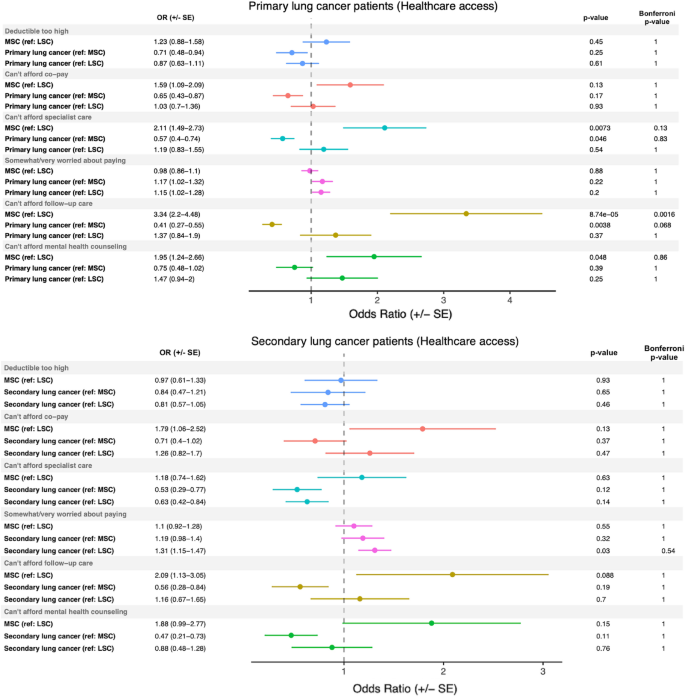
Healthcare access in primary and secondary lung cancer patients. Odds ratios (± standard error) generated comparing healthcare access patient-reported metrics in primary and secondary lung cancer patients to light smoking (LSC) and matched smoking (MSC) controls. The reference group (e.g., (ref: MSC)), mid-p value, and Bonferroni-corrected p-values are reported for each comparison.
Family history patterns in primary and secondary lung cancer
In our investigation of familial history in primary and secondary lung cancer patients, we found that smoking status, rather than lung cancer diagnosis, was associated with an increased odds of having a sibling or father with primary lung cancer (Fig. 2 , top panel). The odds of having a sibling with lung cancer comparing both MSC-1 controls and primary lung cancer patients with LSC-1 controls were 2.31 (p = 0.0020) and 3.17 (p = 4.54e−07), respectively, with both p-values remaining significant after Bonferroni correction. Similarly, the odds of having a father with lung cancer comparing both MSC-1 controls and primary lung cancer patients with LSC-1 controls were 1.66 (p = 0.018) and 1.82 (p = 0.0017), respectively, with the latter maintaining significance after Bonferroni correction. Although the odds of having a mother or grandparent with lung cancer were also increased when comparing our primary lung cancer patients to LSC-1 controls, with odds ratios of 1.75 (p = 0.0087) and 1.74 (p = 0.0083), respectively, neither remained significant after Bonferroni correction. For patients with secondary lung cancer, the odds of having a father with lung cancer were increased with an odds ratio of 1.66 (p = 0.034) compared to LSC-2 controls, while MSC-2 controls compared to LSC-2 controls had an odds ratio of 0.31 (p = 0.00065) of having a grandparent with lung cancer (Fig. 2 , bottom panel).
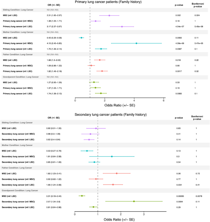
Family history in primary and secondary lung cancer patients. Odds ratios (± standard error) generated comparing family history patient-reported metrics in primary and secondary lung cancer patients to light smoking (LSC) and matched smoking (MSC) controls. The reference group (e.g., (ref: MSC)), mid-p value, and Bonferroni-corrected p-values are reported for each comparison.
Smoking-related behavior in primary and secondary lung cancer
We investigated several smoking-related behaviors, including electronic cigarette use, smokeless tobacco use, hookah use, cigar smoking, and alcohol use, in both primary and secondary lung cancer patients. We observed that primary lung cancer patients had a significantly lower odds of using alcohol compared to all comparison groups (Fig. 3 , top panel). Additionally, primary lung cancer patients had a significantly lower odds of using cigars compared to both MSC-1 and LSC-1 controls, with odds ratios of 0.78 (p = 0.0027) and 0.79 (p = 0.0017), respectively, which retained significance after Bonferroni correction. Interestingly, electronic cigarette use was found to be associated with smoking status rather than lung cancer status, with both primary lung cancer patients and MSC-1 controls having a greater odds of using electronic cigarettes compared to LSC-1 controls, with odds ratios of 3.85 (p = 8.55e−22) and 4.24 (p = 1.46e−22), respectively. These associations also retained significance after Bonferroni correction. Furthermore, primary lung cancer patients demonstrated a nominally significant increased odds of having made a serious smoking quit attempt compared to both MSC-1 and LSC-1 controls, with odds ratios of 1.44 (p = 0.028) and 1.42 (p = 0.026), respectively.
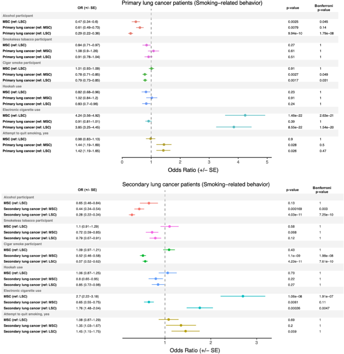
Smoking-related behaviors in primary and secondary lung cancer patients. Odds ratios (± standard error) generated comparing smoking-related behaviors from patient-reported metrics in primary and secondary lung cancer patients to light smoking (LSC) and matched smoking (MSC) controls. The reference group (e.g., (ref: MSC)), mid-p value, and Bonferroni-corrected p-values are reported for each comparison.
In the analysis of smoking-related behaviors in secondary lung cancer patients, both comparison groups had a Bonferroni-corrected significantly lower odds of using alcohol (Fig. 3 , bottom panel). When compared to both MSC-2 and LSC-2 controls, secondary lung cancer patients demonstrated a Bonferroni-corrected significantly lower odds of using cigars, with odds ratios of 0.52 (p = 1.1e−09) and 0.57 (p = 4.23e−11), respectively. Electronic cigarette use was associated with smoking status, rather than lung cancer status. MSC-2 controls had a 2.70 greater odds (p = 1.06e−08) than LSC-2 controls, and secondary lung cancer patients had a 1.76 greater odds (p = 0.00026) than LSC-2 controls of using electronic cigarettes. These associations retained significance after Bonferroni correction.
Primary and secondary lung cancer are associated with significant psychiatric comorbidities
We investigated the odds of lung cancer patients having psychiatric conditions in their electronic health record (EHR) compared to their controls. The analyzed conditions included anxiety, bipolar disorder, depressive disorders, disorders caused by alcohol, insomnia, schizophrenia, and substance use disorder. We found that primary lung cancer patients had significantly higher odds of having substance use disorder, insomnia, bipolar disorder, disorder caused by alcohol, depressive disorder, and anxiety compared to their LSC-1 controls. Each of these odds ratios (except for bipolar disorder) remained significant after Bonferroni correction (Fig. 4 , top panel). MSC-1 controls had significantly greater odds of having a substance use disorder, bipolar disorder, disorder caused by alcohol, anxiety, or a depressive disorder when compared to LSC-1 controls. Furthermore, primary lung cancer patients had significantly higher odds of having anxiety compared to MSC-1 controls (OR: 1.39; p = 0.00052). Interestingly, smoking status was associated with comorbid substance use disorder, bipolar disorder, disorder caused by alcohol, and depressive disorder, instead of primary lung cancer status. Both MSC-1 controls versus LSC-1 controls and primary lung cancer versus LSC-1 controls had a greater odds of having these psychiatric comorbidities. Additionally, secondary lung cancer patients had significantly higher odds of having substance use disorder, insomnia, and anxiety compared to their LSC-2 controls, and these odds ratios retained significance after Bonferroni multiple comparisons adjustment (Fig. 4 , bottom panel). Furthermore, secondary lung cancer patients versus the MSC-2 controls had significantly higher odds of having comorbid insomnia and anxiety.
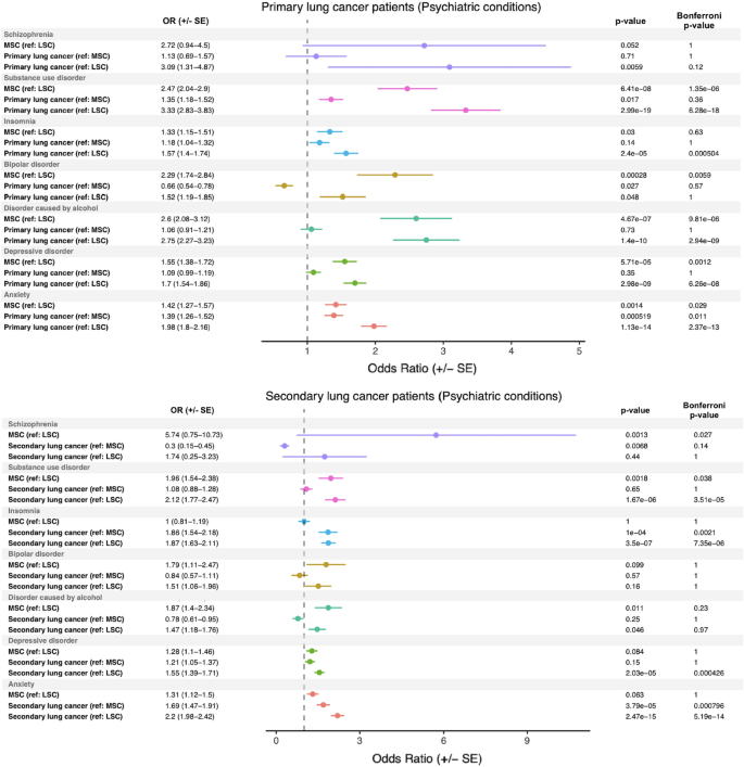
Psychiatric disease burden in primary and secondary lung cancer patients. Odds ratios (± standard error) generated comparing psychiatric disease burden from patient EHR data in primary and secondary lung cancer patients to light smoking (LSC) and matched smoking (MSC) controls. The reference group (e.g., (ref: MSC)), Fisher p-value, and Bonferroni-corrected p-values are reported for each comparison.
In this cross-sectional, case–control study, we examined various epidemiological factors and psychiatric comorbidities in primary and secondary lung cancer. Previous case–control studies on primary lung cancer have investigated factors such as diet 29 , 30 , occupational exposure 31 , 32 , physical activity 33 , medications 34 , 35 , cannabis use 36 , genetic polymorphisms 37 , and various other factors. However, our study is the first to report key epidemiological information in lung cancer from the All of Us Research Program, which has a focus on recruiting historically underrepresented individuals 38 . Additionally, our dual control study design allowed us to differentiate the effect of smoking from the effect of lung cancer when examining variables of interest (Fig. 5 ). We investigated differences in healthcare access, family history, smoking-related behavior, and psychiatric disease burden in our cohort of primary and secondary lung cancer, as well as in the light smoking and matched smoking controls.

Dual control study design. Schema depicting the dual control design utilized in the present study.
The issue of healthcare access and equity is of significant concern in cancer research. In a previous study, it was discovered that cancer death rates in men and women are 13% and 3% higher, respectively, in poorer counties compared to more affluent counties 39 . Furthermore, the same study found that non-Hispanic whites have higher 5-year cancer survival rates than African American, American Indian/Alaskan Native, and Asian/Pacific Islander men 39 . These findings underscore the need to identify and remove barriers to healthcare. Our study produced positive results, as none of the examined lung cancer groups reached Bonferroni-corrected levels of significance for access to care metrics such as increased worry about payment or concern about high copays or deductibles (Fig. 1 ). While our analysis was conducted on the entire cohort of primary and secondary lung cancer patients, future studies can stratify by race, ethnicity, and income to identify potential nuanced differences between these groups regarding access to care metrics.
Family history in primary lung cancer plays an important, yet not fully characterized, role in determining a patient’s predisposition to primary lung cancer 40 . Presently, our results demonstrate that while a first-degree relative with primary lung cancer can increase the odds of a patient having primary lung cancer (Fig. 2 , top panel), family history understandably cannot explain the entire risk. Interestingly, we also saw that an increased odds of having a sibling or father with lung cancer was associated more with smoking behavior, with both primary lung cancer patients and their matched smoking controls compared to light smoking controls having a similar odds of having a sibling or father with lung cancer. This suggests a strong role for the environment in the development of lung cancer, and various literature demonstrates that smoking behavior is correlated in families 41 , 42 , 43 . One major limitation of our family history analysis, however, is that the data is self-reported through a survey, meaning (1) there was no stratification between primary and secondary lung cancer in relatives and (2) not all cases of familial lung cancer will be captured.
In addition to cigarette smoking, we investigated other smoking-related behaviors, such as alcohol use 44 , electronic cigarette use 14 , cigar smoking, hookah use, and smokeless tobacco use. Both primary and secondary lung cancer patients showed a lower odds of using alcohol or cigars, and secondary lung cancer patients also showed a lower odds of using smokeless tobacco compared to the MSC-2 control (Fig. 3 ). Interestingly, in our primary lung cancer patient analysis, electronic cigarette use was associated with smoking status, irrespective of whether or not the patient had primary lung cancer. Namely, smokers (with and without lung cancer) were more likely to use electronic cigarettes compared to their light smoking counterparts. Electronic cigarette use (i.e., vaping) has increased significantly in recent years, and smokers may view vaping as a safer alternative, which can explain the trend observed in this study 14 , 45 . The safety of vaping is under active investigation, and many researchers are concerned about the rapid rise in patients presenting with e-cigarette use-associated lung injury (EVALI) 45 . While vaping may be a more benign alternative to smoking, evidence strongly suggests that vaping has its own associated risks.
Finally, the presence of significant psychiatric comorbidities is well-known in cancer 46 , including lung cancer 47 . By querying electronic health records, we wanted to understand whether or not smoking and/or lung cancer increased the odds of having a comorbid psychiatric condition and by how much. The results from this analysis demonstrated that primary lung cancer patients have a significantly higher odds of having comorbid substance use disorder, insomnia, bipolar disorder, disorder caused by alcohol, depressive disorder, and anxiety when compared to their LSC-1 controls. Secondary lung cancer patients had a significantly higher odds of having substance use disorder, insomnia, and anxiety compared to their LSC-2 controls. However, smoking, rather than lung cancer, appeared to be associated with an increased odds of particular psychiatric comorbidities, such as substance use disorder, bipolar disorder, disorder caused by alcohol, and depressive disorder in primary lung cancer patients. This suggests that much of the psychiatric burden associated with lung cancer may be due to smoking status, rather than lung cancer diagnosis. Of the studied conditions, only secondary lung cancer patients versus matched smoking controls demonstrated a significantly increased odds of comorbid anxiety and insomnia conditions. Additionally, for these two conditions in secondary lung cancer, no significant differences were seen between the matched and light smoking controls, suggesting that the increase in odds was due to secondary lung cancer. Psychiatric conditions like anxiety and depression are well-documented in primary lung cancer 47 , and studies have demonstrated that the mood and anxiety symptoms in lung cancer patients may exceed those of other cancer patients as result of negative psychosocial and physical (e.g., symptom-related) factors 48 . Additionally, perceived negative stigma surrounding primary lung cancer, which is correlated with depressive and anxious symptoms, has been associated with greater psychiatric symptom severity 48 . Furthermore, lung cancer patients may have impaired pulmonary function, leading to lower quality of life (QoL) and increased psychiatric symptom severity 48 , 49 . Understanding the relationship between lung cancer, smoking, and psychiatric disease may help oncologists collaborate closely with mental health professionals to provide well-rounded, comprehensive care to lung cancer patients.
This study did have several limitations, primarily related to known challenges that occur with extracting data from electronic health records and patient surveys. First, in this study, some patients had EHR codes for both primary and secondary lung cancer, leading to an overlap between the primary and secondary lung cancer cohorts of ~ 300 patients. Another challenge is that secondary lung cancer had fewer smokers and fewer patients who filled out the survey indicating the number of years smoked, making it more challenging to assign a full suite of matched smoking controls. Notably, self-reported data from patient surveys may also be subject to bias or inaccuracies. Not every patient in the All of Us database has EMR data, and not all patients who have consented to provide their EMR data have all of their EMR data successfully integrated into the All of Us database, meaning there is a potential for errors or inconsistencies in the coding and categorization of EHR data. Given the size and diversity of the All of Us data network, there are several obstacles related to data integration, and the All of Us team has implemented data quality tools to regularly evaluate, quantify, and communicate about EHR data quality issues 50 . The present study’s cross-sectional design prevents us from making conclusions that establish a temporal relationship between smoking or lung cancer diagnosis and the diagnosis of a comorbid psychiatric condition. Moreover, there is a potential for confounding variables that were not included in the analysis, such as environmental exposures or other health conditions, and the limited number of variables included in the analysis may not fully capture the complex interactions between various epidemiological and clinical factors in lung cancer development and outcomes. Finally, the study’s focus on a specific population may not be representative of the general population, and this analysis should be repeated as the All of Us research program recruits more participants. Of particular note, 60% of participants in the All of Us v6 data release who completed the Basics survey identified as female.
In conclusion, our present cross-sectional, case–control study characterizes primary and secondary lung cancer in the All of Us database, providing information on demographics, healthcare access, family history, smoking-related behaviors, and psychiatric conditions. In future studies, using the vast array of data, including genetic information, present in the All of Us database, researchers can investigate deeper questions, such as probing the combined effect of genetic, environmental, clinical, and epidemiological factors on the development of lung cancer. Future studies can combine genetic models (e.g., polygenic risk scores) with models built from EHR information to improve predictions of disease development, progression, and management, and the All of Us database will be an excellent tool to help researchers answer a wide range of important questions.
Data availability
Data from this program are accessible at http://www.allofus.nih.gov , and this study was conducted on version 6 of the data utilizing the All of Us Researcher Workbench.
Siegel, R. L., Miller, K. D., Fuchs, H. E. & Jemal, A. Cancer statistics, 2022. CA Cancer J. Clin. 72 (1), 7–33. https://doi.org/10.3322/caac.21708 (2022).
Article PubMed Google Scholar
Medeiros, B. & Allan, A. L. Molecular mechanisms of breast cancer metastasis to the lung: clinical and experimental perspectives. Int. J. Mol. Sci. 20 (9), 2272. https://doi.org/10.3390/ijms20092272 (2019).
Article CAS PubMed PubMed Central Google Scholar
Riihimäki, M., Hemminki, A., Sundquist, J. & Hemminki, K. Patterns of metastasis in colon and rectal cancer. Sci. Rep. 6 , 29765. https://doi.org/10.1038/srep29765 (2016).
Article ADS CAS PubMed PubMed Central Google Scholar
Dudani, S. et al. Evaluation of clear cell, papillary, and chromophobe renal cell carcinoma metastasis sites and association with survival. JAMA Netw. Open 4 (1), e2021869. https://doi.org/10.1001/jamanetworkopen.2020.21869 (2021).
Article PubMed PubMed Central Google Scholar
Bozkurt, M., Aghalarov, S., Atci, M. M., Selvi, O. & Canat, H. L. A new biomarker for lung metastasis in non-seminomatous testicular cancer: De Ritis Ratio. Aktuelle Urol 53 (6), 540–544. https://doi.org/10.1055/a-1926-9698 (2022).
Tsuyoshi, H. & Yoshida, Y. Molecular biomarkers for uterine leiomyosarcoma and endometrial stromal sarcoma. Cancer Sci. 109 (6), 1743–1752. https://doi.org/10.1111/cas.13613 (2018).
Islami, F. et al. Proportion and number of cancer cases and deaths attributable to potentially modifiable risk factors in the United States. CA Cancer J. Clin. 68 (1), 31–54. https://doi.org/10.3322/caac.21440 (2018).
Zhou, W. et al. Causal relationships between body mass index, smoking and lung cancer: Univariable and multivariable Mendelian randomization. Int. J. Cancer 148 (5), 1077–1086. https://doi.org/10.1002/ijc.33292 (2021).
Article CAS PubMed Google Scholar
Christiani, D. A. C. et al. Lung cancer: Epidemiology, chapter 74. In Murray and Neidel’s Textbook of Respiratory Medicine 1018–1028 (Elsevier, 2021).
Google Scholar
Schwartz, A. G. & Cote, M. L. Epidemiology of lung cancer. Adv. Exp. Med. Biol. 893 , 21–41. https://doi.org/10.1007/978-3-319-24223-1_2 (2016).
Haddad, D. N., Sandler, K. L., Henderson, L. M., Rivera, M. P. & Aldrich, M. C. Disparities in lung cancer screening: A review. Ann. Am. Thorac. Soc. 17 (4), 399–405. https://doi.org/10.1513/AnnalsATS.201907-556CME (2020).
Bernstein, E., Bade, B. C., Akgün, K. M., Rose, M. G. & Cain, H. C. Barriers and facilitators to lung cancer screening and follow-up. Semin. Oncol. https://doi.org/10.1053/j.seminoncol.2022.07.004 (2022).
Hartmann-Boyce, J., Chepkin, S. C., Ye, W., Bullen, C. & Lancaster, T. Nicotine replacement therapy versus control for smoking cessation. Cochrane Database Syst. Rev. 5 (5), CD000146. https://doi.org/10.1002/14651858.CD000146.pub5 (2018).
Bracken-Clarke, D. et al. Vaping and lung cancer—A review of current data and recommendations. Lung Cancer 153 , 11–20. https://doi.org/10.1016/j.lungcan.2020.12.030 (2021).
Soneji, S. S., Sung, H. Y., Primack, B. A., Pierce, J. P. & Sargent, J. D. Quantifying population-level health benefits and harms of e-cigarette use in the United States. PLoS One 13 (3), e0193328. https://doi.org/10.1371/journal.pone.0193328 (2018).
Coughlin, L. N. et al. Cigarette smoking rates among veterans: Association with rurality and psychiatric disorders. Addict. Behav. 90 , 119–123. https://doi.org/10.1016/j.addbeh.2018.10.034 (2019).
Zhuo, C., Zhuang, H., Gao, X. & Triplett, P. T. Lung cancer incidence in patients with schizophrenia: Meta-analysis. Br. J. Psychiatry 215 (6), 704–711. https://doi.org/10.1192/bjp.2019.23 (2019).
Sikjær, M. G., Løkke, A. & Hilberg, O. The influence of psychiatric disorders on the course of lung cancer, chronic obstructive pulmonary disease and tuberculosis. Respir. Med. 135 , 35–41. https://doi.org/10.1016/j.rmed.2017.12.012 (2018).
Toumazis, I., Bastani, M., Han, S. S. & Plevritis, S. K. Risk-based lung cancer screening: A systematic review. Lung Cancer 147 , 154–186. https://doi.org/10.1016/j.lungcan.2020.07.007 (2020).
Alonso, A. et al. Epidemiology of atrial fibrillation in the All of Us Research Program. PLoS One 17 (3), e0265498. https://doi.org/10.1371/journal.pone.0265498 (2022).
Janjigian, Y. Y. et al. Pack-years of cigarette smoking as a prognostic factor in patients with stage IIIB/IV nonsmall cell lung cancer. Cancer 116 (3), 670–675. https://doi.org/10.1002/cncr.24813 (2010).
Schane, R. E., Ling, P. M. & Glantz, S. A. Health effects of light and intermittent smoking: A review. Circulation 121 (13), 1518–1522. https://doi.org/10.1161/CIRCULATIONAHA.109.904235 (2010).
Husten, C. G. How should we define light or intermittent smoking? Does it matter?. Nicotine Tob. Res. 11 (2), 111–121. https://doi.org/10.1093/ntr/ntp010 (2009).
Aragon, T. epitools: Epidemiology Tools R package version 0.5-10.1. https://CRANR-project.org/package=epitools (2020) (published online) .
Wickham, H. et al. Welcome to the Tidyverse. J. Open Source Softw. 4 (43), 1686. https://doi.org/10.21105/joss.01686 (2019).
Article ADS Google Scholar
Pedersen, T. patchwork: The Composer of Plots R package version 1.1.2. https://CRANR-project.org/package=patchwork (2022) (published online) .
Wickham, H. Ggplot2 (Springer, 2009). https://doi.org/10.1007/978-0-387-98141-3 .
Book MATH Google Scholar
Torre, L. A., Siegel, R. L. & Jemal, A. Lung cancer statistics. Adv. Exp. Med. Biol. 893 , 1–19. https://doi.org/10.1007/978-3-319-24223-1_1 (2016).
Sadeghi, A. et al. Inflammatory potential of diet and odds of lung cancer: A case–control study. Nutr. Cancer 74 (8), 2859–2867. https://doi.org/10.1080/01635581.2022.2036770 (2022).
Krusinska, B. et al. Associations of Mediterranean diet and a posteriori derived dietary patterns with breast and lung cancer risk: A case–control study. Nutrients 10 (4), 470. https://doi.org/10.3390/nu10040470 (2018).
Austin, H., Delzell, E., Lally, C., Rotimi, C. & Oestenstad, K. A case–control study of lung cancer at a foundry and two engine plants. Am. J. Ind. Med. 31 (4), 414–421. https://doi.org/10.1002/(sici)1097-0274(199704)31:4%3c414::aid-ajim6%3e3.0.co;2-v (1997).
Hosseini, B. et al. Lung cancer risk in relation to jobs held in a nationwide case–control study in Iran. Occup. Environ. Med. 79 (12), 831–838. https://doi.org/10.1136/oemed-2022-108463 (2022).
Brizio, M. L. R., Hallal, P. C., Lee, I. M. & Domingues, M. R. Physical activity and lung cancer: A case–control study in Brazil. J. Phys. Act. Health 13 (3), 257–261. https://doi.org/10.1123/jpah.2014-0571 (2016).
Suissa, S., Dell’aniello, S., Vahey, S. & Renoux, C. Time-window bias in case–control studies: Statins and lung cancer. Epidemiology 22 (2), 228–231. https://doi.org/10.1097/EDE.0b013e3182093a0f (2011).
Kristensen, K. B., Hicks, B., Azoulay, L. & Pottegård, A. Use of ACE (angiotensin-converting enzyme) inhibitors and risk of lung cancer: A nationwide nested case–control study. Circ. Cardiovasc. Qual. Outcomes 14 (1), e006687. https://doi.org/10.1161/CIRCOUTCOMES.120.006687 (2021).
Aldington, S. et al. Cannabis use and risk of lung cancer: A case–control study. Eur. Respir. J. 31 (2), 280–286. https://doi.org/10.1183/09031936.00065707 (2008).
Ji, Y., Yang, Y. & Yin, Z. Polymorphisms in lncRNA CCAT1 on the susceptibility of lung cancer in a Chinese northeast population: A case–control study. Cancer Med. 12 (1), 500–512. https://doi.org/10.1002/cam4.4902 (2023).
Denny, J. C. et al. The “All of Us” Research Program. N. Engl. J. Med. 381 (7), 668–676. https://doi.org/10.1056/NEJMsr1809937 (2019).
Ward, E. et al. Cancer disparities by race/ethnicity and socioeconomic status. CA Cancer J. Clin. 54 (2), 78–93. https://doi.org/10.3322/canjclin.54.2.78 (2004).
Dragani, T. A., Manenti, G. & Pierotti, M. A. Polygenic inheritance of predisposition to lung cancer. Ann. Ist. Super. Sanita 32 (1), 145–150 (1996).
CAS PubMed Google Scholar
Joung, M. J., Han, M. A., Park, J. & Ryu, S. Y. Association between family and friend smoking status and adolescent smoking behavior and e-cigarette use in Korea. Int. J. Environ. Res. Public Health https://doi.org/10.3390/ijerph13121183 (2016).
Vàzquez-Nava, F. et al. Association between family structure, parental smoking, friends who smoke, and smoking behavior in adolescents with asthma. ScientificWorldJournal 10 , 62–69. https://doi.org/10.1100/tsw.2010.10 (2010).
Schuck, K., Otten, R., Engels, R. C. M. E., Barker, E. D. & Kleinjan, M. Bidirectional influences between parents and children in smoking behavior: A longitudinal full-family model. Nicotine Tob. Res. 15 (1), 44–51. https://doi.org/10.1093/ntr/nts082 (2013).
DiFranza, J. R. & Guerrera, M. P. Alcoholism and smoking. J. Stud. Alcohol 51 (2), 130–135. https://doi.org/10.15288/jsa.1990.51.130 (1990).
Smith, M. L., Gotway, M. B., Crotty Alexander, L. E. & Hariri, L. P. Vaping-related lung injury. Virchows Arch. 478 (1), 81–88. https://doi.org/10.1007/s00428-020-02943-0 (2021).
Quante, A., Schulz, K. & Fissler, M. Psychiatric comorbidities in cancer patients: Acute interventions by the psychiatric consultation liaison service. Wien Med. Wochenschr. 170 (13–14), 348–356. https://doi.org/10.1007/s10354-020-00739-0 (2020).
Chen, H. M., Tsai, C. M., Wu, Y. C., Lin, K. C. & Lin, C. C. Randomised controlled trial on the effectiveness of home-based walking exercise on anxiety, depression and cancer-related symptoms in patients with lung cancer. Br. J. Cancer 112 (3), 438–445. https://doi.org/10.1038/bjc.2014.612 (2015).
Morrison, E. J. et al. Emotional problems, quality of life, and symptom burden in patients with lung cancer. Clin. Lung Cancer 18 (5), 497–503. https://doi.org/10.1016/j.cllc.2017.02.008 (2017).
Sterzi, S. et al. How best to assess the quality of life in long-term survivors after surgery for NSCLC? Comparison between clinical predictors and questionnaire scores. Clin. Lung Cancer 14 (1), 78–87. https://doi.org/10.1016/j.cllc.2012.04.002 (2013).
Engel, N. et al. EHR data quality assessment tools and issue reporting workflows for the “All of Us” Research Program clinical data research network. AMIA Annu. Symp. Proc. 2022 , 186–195 (2022).
Download references
Acknowledgements
The All of Us Research Program is supported by the National Institutes of Health, Office of the Director: Regional Medical Centers: 1 OT2 OD026549; 1 OT2 OD026554; 1 OT2 OD026557; 1 OT2 OD026556; 1 OT2 OD026550; 1 OT2 OD 026552; 1 OT2 OD026553; 1 OT2 OD026548; 1 OT2 OD026551; 1 OT2 OD026555; IAA #: AOD 16037; Federally Qualified Health Centers: HHSN 263201600085U; Data and Research Center: 5 U2C OD023196; Biobank: 1 U24 OD023121; The Participant Center: U24 OD023176; Participant Technology Systems Center: 1 U24 OD023163; Communications and Engagement: 3 OT2 OD023205; 3 OT2 OD023206; and Community Partners: 1 OT2 OD025277; 3 OT2 OD025315; 1 OT2 OD025337; 1 OT2 OD025276. In addition, the All of Us Research Program would not be possible without the partnership of its participants. V.S. and R.P. would like to thank the Baylor College of Medicine Medical Scientist M.D./Ph.D. training program for their support.
Our study was supported by the National Institutes of Health (NIH) for Integrative Analysis of Lung Cancer Etiology and Risk (U19CA203654) and Sequencing Familial Lung Cancer (R01CA243483). C.I.A. is a Research Scholar of the Cancer Prevention Research Interest of Texas (CPRIT) award (RR170048).
Author information
Authors and affiliations.
Institute for Clinical and Translational Research, Baylor College of Medicine, One Baylor Plaza, Houston, TX, 77030, USA
Vikram R. Shaw, Jinyoung Byun, Rowland W. Pettit, Younghun Han & Christopher I. Amos
Section of Epidemiology and Population Sciences, Department of Medicine, Baylor College of Medicine, Houston, TX, USA
Jinyoung Byun, Younghun Han & Christopher I. Amos
Dan L Duncan Comprehensive Cancer Center, Baylor College of Medicine, Houston, TX, USA
Jinyoung Byun & Christopher I. Amos
School of Medicine, Baylor College of Medicine, Houston, TX, USA
David A. Hsiou & Luke A. Nordstrom
You can also search for this author in PubMed Google Scholar
Contributions
V.S., J.B., R.P., D.H., and L.N. designed the study and analyzed the data. V.S. carried out the implementation. V.S. wrote the manuscript with input from all authors. J.B., Y.H., and C.A. were in charge of overall direction and planning.
Corresponding author
Correspondence to Christopher I. Amos .
Ethics declarations
Competing interests.
The authors declare no competing interests.
Additional information
Publisher's note.
Springer Nature remains neutral with regard to jurisdictional claims in published maps and institutional affiliations.
Supplementary Information
Supplementary information., rights and permissions.
Open Access This article is licensed under a Creative Commons Attribution 4.0 International License, which permits use, sharing, adaptation, distribution and reproduction in any medium or format, as long as you give appropriate credit to the original author(s) and the source, provide a link to the Creative Commons licence, and indicate if changes were made. The images or other third party material in this article are included in the article's Creative Commons licence, unless indicated otherwise in a credit line to the material. If material is not included in the article's Creative Commons licence and your intended use is not permitted by statutory regulation or exceeds the permitted use, you will need to obtain permission directly from the copyright holder. To view a copy of this licence, visit http://creativecommons.org/licenses/by/4.0/ .
Reprints and permissions
About this article
Cite this article.
Shaw, V.R., Byun, J., Pettit, R.W. et al. A comprehensive analysis of lung cancer highlighting epidemiological factors and psychiatric comorbidities from the All of Us Research Program. Sci Rep 13 , 10852 (2023). https://doi.org/10.1038/s41598-023-37585-0
Download citation
Received : 13 April 2023
Accepted : 23 June 2023
Published : 05 July 2023
DOI : https://doi.org/10.1038/s41598-023-37585-0
Share this article
Anyone you share the following link with will be able to read this content:
Sorry, a shareable link is not currently available for this article.
Provided by the Springer Nature SharedIt content-sharing initiative
By submitting a comment you agree to abide by our Terms and Community Guidelines . If you find something abusive or that does not comply with our terms or guidelines please flag it as inappropriate.
Quick links
- Explore articles by subject
- Guide to authors
- Editorial policies
Sign up for the Nature Briefing: Cancer newsletter — what matters in cancer research, free to your inbox weekly.
An official website of the United States government
The .gov means it’s official. Federal government websites often end in .gov or .mil. Before sharing sensitive information, make sure you’re on a federal government site.
The site is secure. The https:// ensures that you are connecting to the official website and that any information you provide is encrypted and transmitted securely.
- Publications
- Account settings
Preview improvements coming to the PMC website in October 2024. Learn More or Try it out now .
- Advanced Search
- Journal List
- Mol Clin Oncol
- v.5(1); 2016 Jul
A case of primary non-small cell lung cancer with synchronous small cell lung cancer
Department of Respiratory Disease, Yijishan Hospital of Wannan Medical College, Wuhu, Anhui 241001, P.R. China
Synchronous multiple primary lung cancer is an uncommon and difficult to distinguish from metastatic disease. The present study reported an extremely rare case of a 66-year-old male with non-small lung cell cancer in the left lobe and synchronous small cell lung cancer in the right lobe. The diagnosis of multiple primary lung cancer not only depends on biopsy pathology, but also requires molecular biology results. This is of great significance for the management and prognosis of multiple primary lung cancer. The management of patients with non-small cell lung cancer-small cell lung cancer produces certain unique challenges, which may require individualized treatment modality that may not strictly comply with standard practices in the setting of a single tumor.
Introduction
Lung cancer is the most common cause of cancer-associated mortality worldwide ( 1 ). Multiple primary lung cancer (MPLC) is rare ( 2 ), ranging between 0.2 and 20% ( 3 ), which has ≥2 primary lung cancers with different pathological types ( 4 ). Two manifestations of MPLC exist: Metachronous or synchronous, and depending on which, their morphology and histology are similar ( 5 ). To distinguish MPLC from metastatic cancer in the lung or recurrence is difficult, however, it is of great significance for therapeutic management and prognosis. The diagnosis of MPLC not only depends on biopsy pathology, but also often requires molecular biology techniques, including DNA polity, gene mutations and microsatellite alteration ( 6 – 9 ). The present study reported a case of primary non-small cell lung cancer (NSCLC) in the left lobe and synchronous small cell lung cancer (SCLC) in the right lung lobe. The patient with synchronous MPLC tolerated two cycles of a chemotherapy regimen that consisted of etoposide (100 mg/m 2 of body-surface area) from one to three days and nedaplatin (75 mg/m 2 of body-surface area) on day one. The patient exhibited a favorable response, including loss of the dry cough and a reduction in the two lesions, observed by chest computed tomography (CT) during follow-up.
A 66-years-old male was referred to the Department of Respiratory Disease at Yijishan Hospital of Wannan Medical College (Wuhu, China) in April 2015 with a dry cough accompanied with blood in phlegm over the previous 1 month. The previous medical history of the patient was uneventful, with the exception of a 40 pack/year history of smoking. Physical examination revealed a rough respiratory murmur in each lung. Laboratory findings were within normal limits, with the exception of prostate special antigen (PSA) at 5.35 ng/ml. Chest CT images obtained in April 2015 revealed an irregular soft-tissue mass with a 9.6 cm maximum diameter, internal uniformity density in the middle-low right lobe close to pulmonary hilum, and a nodular high-density shadow with a 2.5 cm maximum diameter that was a lobulated lesion in the left upper lobe ( Fig. 1A and B ). The patient was positive for metastasis to the mediastinal lymph node. Fiber bronchoscopic biopsy was performed a few days later, which revealed squamous cell carcinoma in the left lobe and SCLC in the right lobe ( Fig. 2 ). Immunohistochemical staining of SCLC revealed a marked positivity for AE1/AE3, Syn, cluster of differentiation 56, thyroid transcription factor-1, p63 (few), Ki-67 (80%) and EMA. By contrast, the SCLC was negative for CgA(−), NapsinA and p40. The patient refused gene detection of squamous cell carcinoma in the left lobe due to expense. The patient tolerated two cycles of a chemotherapy regimen that consisted of etoposide (100 mg/m 2 of body-surface area) from 1–3 days and nedaplatin (75 mg/m 2 of body-surface area) on day 1. The patient exhibited a favorable response, including reduction in the dry cough, and reduction of the two lesions in the chest CT performed during follow-up ( Fig. 1C and D ). A limitation of the present case report is that only two cycles of etopiside combined with nedaplatin were administered in May and June 2015. The treatment with chemotherapy was interrupted due to the large expenditure.

Chest CT scans performed at different stages. (A and B) A chest CT scan performed in April 2015 showing a 96 mm tumor in the middle-low right lobe, and a 25 mm tumor in the left upper lobe, multiple mediastinal lymph nodes, mediastinal and lung windows. (C and D) A chest CT scan performed in June 2015 revealed a 28 mm tumor in the middle-low right lobe, and a patchy tumor of unknown size, multiple mediastinal lymph nodes, mediastinal and lung windows. CT, computed tomography.

Fiber bronchoscopy and biopsy pathology. (A) A new lesion was observed in the left upper lobe. (B) Hematoxylin and eosin staining revealed squamous cell carcinoma in the left lobe. (C) A new lesion was observed in the right middle lobe. (D) Hematoxylin and eosin staining revealed small cell cancer in the right lobe.
The present study reported an uncommon case of a 66-year-old male patient diagnosed with MPLC. Fiber bronchoscopy pathological biopsy revealed two completely different pathological types, which were squamous cell lung carcinoma in the left lobe and SCLC in the right lobe. The patient tolerated two cycles of a chemotherapy regimen that consisted of etoposide (100 mg/m 2 of body-surface area) from 1–3 days and nedaplatin (75 mg/m 2 of body-surface area) on day 1. The patient had a favorable response, including loss of the dry cough and reduction in the two lesions observed by chest CT during the follow-up.
To the best of our knowledge, MPLC is a special type of primary lung carcinoma occurring in one or both lung lobes, which may be ≥2 different pathological types ( 4 ). Two manifestations in MPLC exist: Metachronous or synchronous, and depending on which, their morphology and histology are similar ( 5 ). The mechanism of the MPLC remains to be fully understood, and only a few studies have addressed that it may be associated with field cancerization ( 10 , 11 ). Chang et al ( 12 ) reported the links between MPLC, and the epidermal growth factor receptor (EGFR) and p53 genes. Somatic alterations in EGFR were demonstrated, which can not only greatly improve the clonality assessment, but also affect the management of patients with MPLC. In addition, it is of significance to distinguish MPLC from metastatic carcinoma in the lung for the management and prognosis. The incidence of MPLC is attributed to the development of higher-resolution chest imaging techniques, positron emission computed tomography-CT, fiber bronchoscopy biopsy and percutaneous lung biopsy by CT fluoroscopy. The initial diagnostic criteria were established in 1975 by Martin and Melamed ( 4 ), and were updated by the American College of Chest Physicians (ACCP) ( 13 ). Different pathological categories is of great importance for the identification of MPLC ( 14 ). However, following the Martin-Melamed and ACCP criteria only distinguish certain MPLC types from metastases. Girard et al ( 7 ) confirmed that 3/7 patents with paired adenocarcinoma exhibited multiple primary lung tumors by means of EGFR/KRAS mutation testing. When it comes to similar pathological types, MPLC can be confirmed by means of DNA polity, gene mutations and microsatellite alteration ( 6 – 9 , 15 , 16 ). No standard guidelines exist for the management of MPLC. However, the current case demonstrated a few important principles. SCLC generally exhibits a higher growth fraction, a more rapid doubling time and earlier development of widespread metastases when compared with NSCLC ( 17 ). SCLC is highly sensitive to initial chemotherapy, particularly in the most common regimen (etoposide plus cisplatin), which can provide symptomatic improvement and prolonged survival ( 18 , 19 ). However, Veronesi et al ( 20 ) indicated that outcomes following surgery in patients with early SCLC were comparable to those in patients with NSCLC. The patient with an enlarged mediastinal lymph node exhibited squamous carcinoma combined with SCLC, therefore, that chemotherapy with etoposide and nedaplatin was performed. Therefore, if an MPLC patient is confirmed with synchronous SCLC and NSCLC, the appropriate chemotherapy regimen must be selected as quickly as possible.
In conclusion, the present study reported a case of MPLC with simultaneous SCLC and NSCLC. The management of patients with MPLC requires highly individualized treatment plans that are dissimilar to standard practices in the setting of a single tumor. Consideration must be given to the chemotherapy regimen that improve symptoms and prolongs survival.
Acknowledgements
The authors would like to thank Renguang Pei (Department of Interventional Therapy, Yijishan Hospital of Wannan Medical College, Wuhu, China) for providing assistance with writing and organization of this manuscript.
8 Lung Cancer Nursing Care Plans

Utilize this comprehensive nursing care plan and management guide to deliver effective care for patients with lung cancer . Gain valuable insights on nursing assessment , interventions, goals, and nursing diagnoses specifically tailored for lung cancer in this guide.
Table of Contents
What is lung cancer, nursing problem priorities, nursing assessment, nursing diagnosis, nursing goals, 1. improving gas exchange, 2. managing pain and discomfort, 3. maintaining patent airway clearance, 4. administering medications and pharmacological support, 5. reducing fear and anxiety, 6. promoting optimal nutritional balance, 7. promoting rest and tolerance to activity, 8. providing patient education & health teachings, recommended resources, references and sources.
Lung cancer or bronchogenic carcinoma refers to tumors originating in the lung parenchyma or within the bronchi (Munakomi, 2022). Lung cancers are generally divided into two main categories: small-cell lung cancer (SCLC) and non-small-cell lung cancer (NSCLC). NSCLC accounts for approximately 85% of all lung cancers. It is further divided into adenocarcinoma, squamous cell carcinoma, (SCC), and large cell carcinoma. SCLC is considered distinct from other lung cancers because of its clinical and biological characteristics (Tan & Karim, 2021).
Adenocarcinoma is the most common NSCLC cancer in the United States. It arises from the bronchial mucosal glands, a subtype observed most commonly in persons who do not smoke. Squamous cell carcinoma accounts for 25 to 30% of all lung cancers. SCC is found in the central parts of the lung. It is the type most often associated with hypercalcemia . Large cell carcinoma typically manifests as a large peripheral mass on the chest radiograph. This type accounts for 10 to 15% of lung cancers (Tan & Karim, 2022).
Smoking is the most common cause of lung cancer. It is estimated 90% of lung cancer cases are attributable to smoking. It is interesting to note that lung cancer was a relatively rare disease at the beginning of the 20th century. Its dramatic rise in later decades is mainly attributable to the increase in smoking among males and females (Munakomi, 2022).
No specific signs and symptoms exist for lung cancer. Lung cancer symptoms occur due to local effects of the tumor , such as cough due to bronchial compression by the tumor due to distant metastasis, stroke -like symptoms secondary to brain metastasis, paraneoplastic syndrome, and kidney stones due to persistent hypercalcemia (Munakomi, 2022).
Nursing Care Plans & Management
Nursing care for patients with lung cancer encompasses both physiological and psychological aspects, similar to other cancer patients. The focus is on addressing the respiratory manifestations of the disease, along with providing pain relief, managing discomfort, and preventing complications. Strategies are implemented to meet the patient’s needs and ensure their overall well-being.
The following are the nursing priorities for patients with lung cancer:
- Relieving breathing problems
- Managing symptoms of lung cancer
- Reducing fatigue
- Providing emotional support
- Patient education and health teachings
Assess for the following subjective and objective data :
- Restlessness/changes in mentation
- Hypoxemia and hypercapnia
- Changes in rate/depth of respiration
- Abnormal breath sounds
- Ineffective cough
Assess for factors related to the cause of lung cancer:
- Increased amount/viscosity of secretions
- Restricted chest movement / pain
- Fatigue/ weakness
- Surgical incision, tissue trauma , and disruption of intercostal nerves
- Presence of chest tube(s)
- Cancer invasion of the pleura, chest wall
Following a thorough assessment , a nursing diagnosis is formulated to specifically address the challenges associated with lung cancer based on the nurse ’s clinical judgement and understanding of the patient’s unique health condition. While nursing diagnoses serve as a framework for organizing care, their usefulness may vary in different clinical situations. In real-life clinical settings, it is important to note that the use of specific nursing diagnostic labels may not be as prominent or commonly utilized as other components of the care plan. It is ultimately the nurse’s clinical expertise and judgment that shape the care plan to meet the unique needs of each patient, prioritizing their health concerns and priorities.
Goals and expected outcomes may include:
- The client will demonstrate improved ventilation and adequate oxygenation of tissues by ABGs within the normal range.
- The client will be free of symptoms of respiratory distress.
- The client will maintain a patent airway with clear breath sounds.
- The client will clear secretions and be free of aspiration .
- The client will report pain relief /control.
- The client will appear relaxed and sleep /rest appropriately.
- The client will participate in desired/needed activities.
Nursing Interventions and Actions
Nursing interventions for patients with lung cancer encompass pain management, respiratory support, symptom management, psychological support, education and health promotion , nutritional support, collaboration and coordination , end-of-life care, and supportive care. These interventions aim to address the physiological and psychological needs of the patients, optimize their comfort, manage symptoms, provide education and support, and enhance their overall well-being throughout their journey with lung cancer. Therapeutic interventions and nursing actions for patients with lung cancer may include:
Centrally located obstructing tumors can cause the collapse of the entire lung with an absence of breath sounds on the side of the lesion. Rapid tumor growth may lead to obstruction of major airways, with distal collapse leading to post-obstructive pneumonia , infection , and fever (Tan & Karim, 2021).
Note respiratory rate, depth, and ease of respiration. Note the use of accessory muscles and pursed-lip breathing . Respirations may be increased as a result of pain or as an initial compensatory mechanism to accommodate for the loss of lung tissue. Additionally, respiratory insufficiency is signaled by dyspnea and increased work of breathing, retractions, orthopnea, and cyanosis. In SCLC, clients usually experience shortness of breath; physical examination may reveal the use of the accessory muscles of respiration and nasal flaring (Tan & Karim, 2021).
Observe changes in skin or mucous membrane color, pallor, cyanosis, and edema . Increased work of breathing and cyanosis may indicate increasing oxygen consumption, energy expenditures, and reduced respiratory reserve. Examination of the extremities may reveal clubbing, cyanosis, or edema . In the presence of superior vena cava (SVC) obstruction, the right upper extremity is usually edematous (Tan & Karim, 2021).
Auscultate lungs for air movement and abnormal breath sounds. Consolidation and lack of air movement on the operative side are normal in the pneumonectomy client; however, the lobectomy client should demonstrate normal airflow in the remaining lobes. In clients diagnosed with NSCLC, upper airway obstruction is manifested by stridor and wheezing. Lower airway obstruction is manifested by asymmetric breath sounds, pleural effusion, pneumothorax , infiltrates, and post-obstructive processes (Tan & Karim, 2022).
Investigate restlessness and changes in mentation or level of consciousness. A neurologic examination should be performed to assess for focal neurological deficits caused by brain metastases and for signs of spinal cord compression (Tan & Karim, 2022). This may also indicate increased hypoxia or complications such as a mediastinal shift in the pneumonectomy client when accompanied by tachypnea , tachycardia, and tracheal deviation.
Assess the client’s response to the activity. Increased oxygen consumption demand and stress of surgery can result in increased dyspnea and changes in vital signs with activity; however, early mobilization is desired to help prevent pulmonary complications and to obtain and maintain respiratory and circulatory efficiency.
Note the development of fever. Fever within the first 24 hours after surgery is frequently due to atelectasis . Temperature elevation within the 5th to 10th postoperative day usually indicates a wound or systemic infection.
Assess for cough and mucus production, hemoptysis, and chest pain . Cough is present in 50 to 75% of clients diagnosed with lung cancer. Cough productive of large volumes of thin, mucoid secretions is seen in mucinous adenocarcinoma. In some cases, especially those with exophytic bronchial masses, a cough may signify secondary post-obstructive pneumonia . Hemoptysis is present in 15 to 30% of clients with lung cancer. Chest pain is present in approximately 20 to 40% of clients (Munakomi, 2022).
Monitor and graph ABGs, and pulse oximetry readings. Note hemoglobin (Hb) levels. Decreasing Pao 2 or increasing Pco2 may indicate the need for ventilatory support. Significant blood loss can result in decreased oxygen-carrying capacity, reducing Pao 2 . Arterial blood gas ( ABG ) levels are useful in the detection of respiratory failure in sick clients. Obtain ABG levels in clients with active systemic diseases or abnormal labored breathing (Tan & Karim, 2022).
Monitor chest radiography results and other imaging tests as indicated. A chest radiograph is usually the first test ordered in clients in whom a lung malignancy is suggested. Clues from the chest radiograph may suggest the diagnosis of lung cancer, but may not be helpful in identifying a histologic subtype. A chest CT scan is a standard for lung cancer staging. The findings of CT scans of the chest and clinical presentation usually allow a presumptive differentiation between NSCLC and SCLC (Tan & Karim, 2022).
Encourage rest periods and limit activities according to client tolerance. Adequate rest balanced with activity can prevent respiratory compromise. The client’s activity level, as measured by a performance status scale is an important prognostic factor. The client should be encouraged to remain active during and after treatment for lung cancer (Tan & Karim, 2022).
Educate regarding smoking cessation. Advise clients that smoking cessation is the most important measure for preventing lung cancer; it may also improve prognosis in clients with early-stage lung cancer. Smoking cessation by others who share the client’s home, car, or both is also important. According to published data, the use of nicotine alternatives instead of cigarettes reduces the incidence of lung cancer, although it does not affect the incidence of ischemic heart disease (Tan & Karim, 2022).
Maintain patent airway by positioning, suctioning, and use of airway adjuncts. Airway obstruction impedes ventilation, impairing gas exchange . In the case of upper airway obstruction, the client is admitted to the ICU, and prepared for intubation and/or cricothyrotomy and intraoperative tracheostomy (Tan & Karim, 2022).
Reposition frequently, placing the client in sitting positions and supine to side positions. However, avoid positioning the client with a pneumonectomy on the operative side; instead, favor the “good lung down” position. This maximizes lung expansion and drainage of secretions. Research shows that positioning clients following lung surgery with their “good lung down” maximizes oxygenation by using gravity to enhance blood flow to the healthy lung, thus creating the best possible match between ventilation and perfusion (Lan et al., 2011).
Encourage and assist with deep-breathing exercises and pursed-lip breathing as appropriate. This promotes maximal ventilation and oxygenation and reduces or prevents atelectasis. Breathing exercises aim to correct breathing errors, reestablish proper breathing patterns, increase diaphragm activity, elevate the amount of alveolar ventilation, reduce energy consumption when breathing, and relieve the shortness of breath experienced by clients diagnosed with lung cancer (Liu et al., 2013).
Maintain patency of the chest drainage system for lobectomy, segmental, or wedge resection. These drain fluid from the pleural cavity to promote the re-expansion of remaining lung segments. The balanced chest drainage system was found to be associated with reduced rates of post-pneumonectomy pulmonary edema , which is a common cause of death after pneumonectomy (Wei Lo et al., 2020).
Note changes in the amount or type of chest tube drainage. Bloody drainage should decrease in amount and change to a more serous composition as recovery progresses. A sudden increase in the amount of bloody drainage or return to frank bleeding suggests thoracic bleeding or hemothorax ; sudden cessation suggests blockage of the tube, requiring further evaluation and intervention.
Observe the presence or degree of bubbling in the water-seal chamber. Air leaks immediately postoperative are not uncommon, especially following lobectomy or segmental resection; however, this should diminish as healing progresses. Most intrathoracic air leaks will usually seal spontaneously, and resolution can be tracked by witnessing decreased bubbling in the device over days. Prolonged or new leaks require evaluation to identify problems in clients versus the drainage system (Merkle & Cindass, 2022).
Administer supplemental oxygen via nasal cannula, partial rebreathing mask, or high-humidity face mask, as indicated. This maximizes available oxygen, especially while ventilation is reduced because of anesthetic, depression, or pain, and during the period of the compensatory physiological shift of circulation to remaining functional alveolar units. If hemoptysis is noted, administer supplemental oxygen and perform suctioning. If a threat of imminent demise exists, consider placing a double-lumen endotracheal tube (Tan & Karim, 2022).
Assist with and encourage the use of incentive spirometer . This prevents or reduces atelectasis and promotes the re-expansion of small airways. Lung expansion therapy allows the client to maintain an effective cough mechanism to facilitate the removal of secretions from the airways following surgery. An incentive spirometer is a medical device, which helps the client sustain maximal inspiration under visual quantitation by inspiratory effort (Liu et al., 2019).
Pain is one of the most prevalent symptoms in clients diagnosed with lung cancer; it can arise from local invasion of chest structures or metastatic disease invading bones , nerves, or other anatomical structures that are potentially painful. Pain can also be a consequence of therapeutic approaches like surgery, chemotherapy , or radiotherapy (Hochberg et al., 2017).
Ask the client about pain. Determine pain characteristics: continuous, aching, stabbing, burning. Have the client rate intensity on a 0–10 scale. Identifying the level of pain is helpful in evaluating cancer-related pain symptoms, which may involve viscera, nerve, or bone tissue. The use of a rating scale aids the client in assessing the level of pain and provides a tool for evaluating the effectiveness of analgesics, enhancing the client’s control of pain. Acute cancer pain is usually due to a definable acute injury or illness. Chronic cancer pain can result from the same causes as acute pain but is differentiated by longevity (Simmons et al., 2012).
Assess the client’s verbal and nonverbal pain cues. The discrepancy between verbal and/or nonverbal cues may provide clues to the degree of pain, need for, or effectiveness of interventions. Acute pain is associated with clinical signs of sympathetic overactivity such as tachycardia, hypertension , sweating, pupillary dilatation, and pallor (Simmons et al., 2012).
Note possible pathophysiological and psychological causes of pain. Fear , distress, anxiety , and grief over the confirmed diagnosis of cancer can impair the ability to cope. In addition, a posterolateral incision is more uncomfortable for the client than an anterolateral incision. The presence of chest tubes can greatly increase discomfort. The three main causes of pain in clients diagnosed with advanced lung cancer are skeletal metastatic disease (34%), Pancoast tumor (31%), and chest wall disease (21%) (Simmons et al., 2012).
Evaluate the effectiveness of pain control. Encourage sufficient medication to manage pain; change medication or time span as appropriate. Pain perception and pain relief are subjective, thus pain management is best left to the client’s discretion. If the client cannot provide verbal input, the nurse should observe physiological and nonverbal signs of pain and administer medications regularly. Undertreated cancer pain associates physical and psychological consequences, causing suffering and reduced quality of life (Hochberg et al., 2017).
Assess the client’s understanding of the evaluation and pain relief strategies. This review helps determine the client’s level of understanding and reinforces findings, thereby promoting knowledge and adherence to pain relief strategies. It also empowers the client as much as possible to participate in controlling their pain.
Assess the client’s cultural beliefs and attitudes about pain. Never ignore a client’s report of pain. Cultural beliefs may influence how individuals describe their pain and its severity and their willingness to ask for pain medications. Pain is dynamic, and competent management requires frequent assessment at scheduled intervals.
Assess the client’s and caregiver ’s attitudes and knowledge about the pain medication regimen. Many clients and their families have fears related to the client’s ultimate addiction to opioids. It is important to dispel any misperceptions about opioid-induced addiction when chronic pain therapy is necessary. Fears of addiction may result in ineffective pain management.
Encourage verbalization of feelings about the pain. Fears or concerns can increase muscle tension and lower the threshold of pain perception. An interesting finding from studies with cancer clients shows a low mean score on the client’s knowledge and attitudes toward cancer pain management. This result may be explained by the fact that many clients could be reluctant to report their pain to professionals because they have a mistaken belief regarding opioid medication (Makhlouf et al., 2019).
Provide comfort measures: frequent changes of position, back rubs, and support with pillows. Encourage the use of relaxation techniques, visualization, guided imagery, and appropriate diversional activities. Nonpharmacological management of pain also promotes relaxation and redirects attention. They may relieve discomfort and augment the therapeutic effects of analgesia. Nonpharmacologic approaches such as acupressure, biofeedback, application of heat or cold, and massage are often effective in enhancing the effects of opioid therapy.
Schedule rest periods, and provide a quiet environment. Providing the client with rest periods decreases fatigue and conserves energy, enhancing coping abilities. A quiet environment decreases external stimuli, which can aggravate anxiety and limit the client’s coping abilities and adjustment to the current situation.
Assist with self-care activities, breathing and/or arm exercises, and ambulation. This prevents undue fatigue and incisional strain. Encouragement and physical assistance and support may be needed for some time before the client is able or confident enough to perform these activities because of pain or fear of pain. Caregivers with a higher pain management knowledge have significantly fewer barriers to cancer pain management, therefore, they should have a general awareness and adequate level of knowledge to be able to assist the client with daily activities (Makhlouf et al., 2019).
Assist with patient-controlled analgesia (PCA) or analgesia through the epidural catheter. Maintaining a constant drug level avoids cyclic periods of pain, aids in muscle healing, and improves respiratory function and emotional comfort, and coping. The goal of PCA is to efficiently deliver pain relief at a client’s preferred dose and schedule by allowing them to administer a predetermined bolus dose of medication on-demand at the press of a button. PCA has proven to be more effective at pain control than non-patient opioid injections and results in higher client satisfaction, especially in clients who are unable to tolerate oral medications (Chen et al., 2022).
Administer intermittent analgesics routinely as indicated, especially 45–60 min before respiratory treatments, deep-breathing, or coughing exercises . The World Health Organization’s (WHO) Analgesic Ladder for Cancer Pain Relief provides a stepwise approach to managing pain in clients diagnosed with cancer. Step 1 advises the use of paracetamol or a nonsteroidal anti-inflammatory drug. If the client is not satisfactorily controlled, it is appropriate to move to Step 2 Analgesia which includes the use of weak opioids, usually codeine . Morphine is the usual first-line Step 3 opioid, however, there are many alternatives to morphine nowadays (Simmons et al., 2012).
Recognize and report/treat side effects of opioid analgesia early. Side effects include respiratory depression, nausea and vomiting , constipation , sedation, and itching. The presence of these side effects does not necessarily preclude continued use of the drug. Consult with the care provider regarding the prophylactic use of stool softeners to prevent constipation.
Avoid stopping opioids abruptly in clients who have been taking them for a prolonged period. There is potential for physical dependence in clients taking opioids for a prolonged period; therefore, they should be tapered gradually to prevent withdrawal discomfort. Morphine, like other strong opioids, is titrated until the desired analgesic benefit is achieved (Simmons et al., 2012).
Educate the client and SOs about complementary therapies for pain management. Complementary therapies are used as adjuncts to current evidence-based management. They are supportive measures that assist in symptom control, enhance well-being, and contribute to overall client care. However, it is important to evaluate herbal and other dietary products for side effects and potential interactions with chemotherapy and other medications (Simmons et al., 2012).
Provide additional information about interventional pain medicine. Interventional pain management is a subspecialty of medicine devoted to the use of invasive techniques such as joint injections, nerve blocks and/or neurolysis, neuromodulation, and cement augmentation techniques to provide diagnosis and treatment of pain syndromes being unresponsive to conventional medical management. Several interventional cancer pain procedures have demonstrated effectiveness in relieving drug-resistant cancer pain symptoms, yet the evidence is scant (Hochberg et al., 2017).
Lung cancer may narrow the airway, causing wheezing. If a tumor blocks an airway, part of the lung that the airway supplies may collapse, a condition called atelectasis. Other consequences of a blocked airway are shortness of breath and pneumonia, which may result in coughing, fever, and chest pain (Keith, 2022).
Auscultate the chest for the character of breath sounds and the presence of secretions. Noisy respirations, rhonchi, and wheezes indicate retained secretions or airway obstruction. Centrally located obstructing tumors can cause the collapse of the entire lung with an absence of breath sounds on the side of the lesion. Upper airway obstruction is manifested by stridor and wheezing. Lower airway obstruction is manifested by asymmetric breath sounds (Tan & Karim, 2022).
Observe the amount and character of sputum or aspirated secretions. Investigate changes as indicated. Increased amounts of colorless, blood-streaked, or watery secretions are normal initially and should decrease as recovery progress. The presence of thick or tenacious, bloody, or purulent sputum suggests the development of secondary problems ( dehydration , pulmonary edema, local hemorrhage , or infection) that require correction and treatment.
Assess for pain or discomfort and medicate on a routine basis and before breathing exercises. Most peripheral tumors are adenocarcinomas or large cell carcinomas and, in addition to causing cough and dyspnea, can cause severe pain as a result of infiltration of the parietal pleura and the chest wall (Tan & Karim, 2022). Pre-medication encourages the client to move, cough more effectively, and breathe more deeply to prevent respiratory insufficiency.
Assess the rate and depth of respirations and chest movement. Dyspnea is prominent in clients early in their disease. Causes of dyspnea may include tumor occlusion of the airway or lung parenchyma, pleural effusion, pneumonia, or complications of treatment. Tachypnea, shallow respirations, and asymmetric chest movements are frequently present because of the discomfort of moving chest wall or fluid in the lung.
Observe for the characteristics of cough. The most frequent symptom of lung cancer is a cough or a change in a chronic cough. People frequently ignore this symptom and attribute it to smoking or a respiratory infection. The cough may start as a dry, persistent cough, without sputum production. When obstruction of the airways occurs, the cough may become productive due to infection.
Monitor serial ABGs and chest X-ray s. Arterial blood gas (ABG) levels are useful in the detection of respiratory failure. It is obtained in clients with active systemic diseases or abnormal labored breathing. A chest radiography is usually the first test ordered in clients in whom a lung malignancy is suggested. Clues from the chest radiograph may suggest the diagnosis of lung cancer. If the tumor is clearly visible and measurable, chest radiography can sometimes be used to monitor response to therapy (Tan & Karim, 2022).
Elevate the client’s head of the bed and change positions frequently. Keeping the head of the bed elevated lowers the diaphragm, promoting chest expansion, aeration of lung segments, and mobilization and expectoration of secretions to keep the airway clear.
Assist the client and instruct effective deep breathing and coughing with an upright position (sitting) and splinting of an incision. Upright position favors maximal lung expansion and splinting improving the force of cough effort to mobilize and remove secretions. Splinting may be done by the nurse (placing hands anteriorly and posteriorly over the chest wall) and by the client (with pillows) as strength improves. In their simplest form, breathing exercises consist of elongating and slowing down the inhalation and exhalation, which allow lung cancer clients to take deeper breaths that increase their intake of oxygen, rather than taking shallow breaths that only make use of the top half of their lungs (Liu et al., 2019).
Suction if cough is weak or breath sounds not cleared by cough effort. Suction the client as needed, and encourage them to begin deep breathing and coughing as soon as possible. “Routine” suctioning increases the risk of hypoxemia and mucosal damage. Deep tracheal suctioning is generally contraindicated following pneumonectomy to reduce the risk of rupture of the bronchial stump suture line. Avoid deep endotracheal or nasotracheal suctioning in pneumonectomy clients if possible. If suctioning is unavoidable, it should be done gently and only to induce effective coughing. Suctioning can stimulate the vagal nerve, predisposing the client to bradycardia and hypoxia. Hypoxia can be profound from occlusion interruption of oxygen supply, and prolonged suctioning (Sinha et al., 2022).
Encourage oral fluid intake (at least 2500 mL/day) within cardiac tolerance. Adequate hydration aids in keeping secretions loose or enhances expectoration. Using warm liquids may decrease bronchospasm. Fluid during meals can increase gastric distention and pressure on the diaphragm.
Assist with an incentive spirometer, postural drainage , and percussion as indicated. These techniques improve lung expansion or ventilation and removal of secretions. Postural drainage may be contraindicated in some clients and in any event, must be performed cautiously to prevent respiratory embarrassment and incisional discomfort. Airway clearance techniques are key to maintaining airway patency through the removal of excess secretions. This may be accomplished through deep-breathing exercises, chest physiotherapy , directed cough, suctioning, and in some instances bronchoscopy .
Use humidified oxygen and/or ultrasonic nebulizer . Provide additional fluids via IV as indicated. Providing maximal hydration helps loosen or liquefy secretions to promote expectoration. Impaired oral intake necessitates IV supplementation to maintain hydration. Oxygen is commonly prescribed for lung cancer clients with advanced disease. Indications include hypoxemia and dyspnea. Reversal of hypoxemia in some cases will alleviate dyspnea (Tiep et al., 2013).
Bronchodilators Bronchodilators relieve bronchospasm to improve airflow. As the tumor enlarges or spreads, it may compress a bronchus or involve a large area of lung tissue, resulting in impaired breathing patterns and poor gas exchange .
Expectorants Expectorants increase mucus production and liquefy and reduce the viscosity of secretions, facilitating removal. They exert their effect on the mucus layer lining the respiratory tract with the motive of enhancing its clearance (Gupta & Wadhwa, 2022).
Analgesics Alleviation of chest discomfort promotes cooperation with breathing exercises and enhances the effectiveness of respiratory therapies. The World Health Organization’s (WHO) Analgesic Ladder for Cancer Pain Relief provides a stepwise approach to managing pain in clients diagnosed with cancer. Step 1 advises the use of paracetamol or a nonsteroidal anti-inflammatory drug. If the pain is not satisfactorily controlled, it is appropriate to move to step 2 analgesia which includes the use of weak opioids, usually codeine . Clients with severe pain usually need step 3 analgesia, the use of strong opioids (Simmons et al., 2012).
Psychological distress is also related to the symptoms of clients diagnosed with cancer and consequently, psychological support and care must be integral to the client’s treatment. Untreated psychological distress may exacerbate pain or other symptoms. It is therefore important to ensure clients have access to counseling and spiritual support (Simmons et al., 2012).
Evaluate the client’s and significant other ‘s (SO) level of understanding of the diagnosis. The client and SO are hearing and assimilating new information that includes changes in self-image and lifestyle. Understanding the perceptions of those involved sets the tone for individualizing care and provides information necessary for choosing appropriate interventions.
Assess the client’s anxiety levels in association with the COVID-19 pandemic . Cancer clients represent an already compromised group known to have elevated levels of fear and anxiety, and the introduction of the pandemic places this vulnerable population at an even higher risk for mental health consequences (Albano et al., 2021).
Assess mental status, including mood and affect, comprehension of events, and content of thoughts, such as illusions or manifestations of terror or panic . Initially, the client may use denial and repression to reduce and filter information that might be overwhelming. Some clients display a calm manner and alert mental status, representing dissociation from reality, which is also a protective mechanism.
Identify previous methods of coping with and handling stressful situations. Past successful behavior can be used to assist in dealing with the present situation. Coping style is considered to be potentially related to psychological distress, and the association between coping style and psychological distress has been demonstrated (Tian et al., 2021).
Acknowledge the reality of the client’s fears or concerns and encourage the expression of feelings. Support may enable the client to begin exploring and dealing with the reality of cancer and its treatment. The client may need time to identify feelings and even more time to begin to express them. Provide an open environment in which the client feels safe to discuss their feelings and help them feel accepted in their present condition. This may promote a sense of dignity and control.
Provide an opportunity for questions and answer them honestly. Be sure that the client and care providers have the same understanding of the terms used. This establishes trust and reduces misperceptions and/or misinterpretation of information Providing the client with accurate and consistent information can reduce anxiety and enable the client to make decisions and choices based on realities.
Accept, but do not reinforce, the client’s denial of the situation. When extreme denial or anxiety is interfering with the progress of recovery, the issues facing the client need to be explained and resolutions explored. Previous studies have shown that women with advanced cancer applied denial or avoidance to cope with their disease. Increasing denial or escape-avoidance coping has been associated with a high level of emotional distress (Liao et al., 2018).
Note comments or behaviors indicative of beginning acceptance and/or use of effective strategies to deal with the situation. Fear and/or anxiety will diminish as the client begins to accept or deal positively with reality. An indicator of the client’s readiness to accept responsibility for participation in recovery and to “resume life.” Evidence indicated that clients with a high level of positive attitude might have more expectations and confidence in addressing adverse challenges and vice versa (Tian et al., 2021).
Involve the client and SO in care planning . Provide time to prepare for events or treatments. This may help restore some feeling of control or independence to the client who feels powerless in dealing with diagnosis and treatment. Social support has been identified as a buffering or protective source of distress and psychosocial adjustment (Tian et al., 2021).
Provide for the client’s physical comfort. It is difficult to deal with emotional issues when experiencing extreme or persistent physical discomfort. In a study, clients and caregivers described physical strategies for coping with anxiety including exercise, meditation, slow breathing, and other forms of focused relaxation . Exercise to alleviate anxiety was usually pursued separately by clients and caregivers due to differing degrees of health and energy, but they also practiced other forms of relaxation together such as meditation and going for walks (Hendriksen et al., 2019).
Identify manifestations of anxiety and coping strategies to alleviate them.
- Cognitive Cognitive manifestations of anxiety are present in both clients and caregivers. Some of the methods used by clients and caregivers in a study include waiting it out using visualization strategies, using mindfulness, or engaging in activities (Hendriksen et al., 2019).
- Behavioral The first behavioral manifestation of anxiety concerns exertion. Some clients respond by becoming behaviorally cautious; they feel fragile or endangered and act in accordance. To cope with these manifestations, clients and caregivers sought support from others. They may connect with friends or can find respite by pursuing activities with others or making use of mental health care resources (Hendriksen et al., 2019).
- Physiological Physiological manifestations and strategies for coping with anxiety are separate from behavioral ones as they focus on the physical body. Clients and caregivers may report impairing physical symptoms of anxiety, from muscle tension to gastrointestinal upset. The most common shared symptom was sleep disruption, experienced by clients and caregivers. They may cope with these manifestations through exercise, meditation, slow breathing, and other forms of focused relaxation (Hendriksen et al., 2019).
Patients with lung cancer often experience nutritional deficiencies, especially those in whom the disease is at an advanced stage and those with metastatic disease. Different aspects contribute to cancer-related malnutrition : inadequate caloric intake, metabolic derangements, depression, fatigue, and chemotherapy-induced toxicity (CIT), leading to a loss of skeletal muscle mass and a systemic inflammation syndrome, in which acute phase proteins are altered (Mele et al., 2020).
Weigh the client regularly. Nausea, vomiting , anorexia, and taste changes all may contribute to weight loss . Weight loss and body changes are frequently experienced by clients during the therapeutic process. Weight loss and a lower BMI significantly worsen post- surgical and survival outcomes (Mele et al., 2020).
Assess food likes and dislikes, as well as cultural and religious preferences related to food choices. Providing foods on the client’s “like” list as often as feasible and avoiding foods on the “dislike” list optimally will promote sufficient intake. However, foods previously enjoyed may become undesirable, whereas previously disliked foods may appeal.
Assess the client’s pattern of nausea and vomiting : onset, frequency, duration, intensity, and the amount and character of emesis. Knowledge about the pattern of nausea and vomiting enables the use of proper medication, route, and timing. Nausea and vomiting are two serious side effects of cancer chemotherapy. These adverse effects can cause significant negative impacts on the client’s quality of life and on their ability to comply with therapy (Mancini, 2012).
Assess skin and mucous membranes for pallor, delayed wound healing , and enlarged parotid glands. This helps in the identification of protein-calorie malnutrition, especially when weight and anthropometric measurements are less than normal.
Explain that anorexia may be caused by the pathophysiology of cancer and surgery or side effects of chemotherapy and radiation therapy. Taste and olfactory receptors have a high rate of cell growth and may be sensitive to chemotherapy and radiation therapy. During the administration of cytotoxic chemotherapy, the chemosensory systems are exposed to more changes than other sensory systems due to the short life span of gustatory and olfactory receptor cells and their frequent renewal (Drareni et al., 2019).
Encourage the client to eat several small meals at frequent intervals throughout the day. Smaller, more frequent meals are usually better tolerated than larger meals. Metabolic tissue and needs are increased to eliminate waste products. Smaller meals decrease client intake, therefore also reducing fatigue during meals. Additionally, presenting large volumes of food can be overwhelming, thereby extinguishing the appetite or causing nausea.
Promote the use of nutritional supplements. Adequate protein and calories are important for healing, fighting infection, and providing energy. Supplements can play an important role in maintaining adequate caloric and protein intake. Several studies demonstrated the efficacy of oral nutritional supplements, especially those enriched with fatty acids, given their capability to modulate the pro-inflammatory cascade (Mele et al., 2020).
Encourage increased intake of calorie-rich and protein-rich foods. Increasing calories augment energy, minimize weight loss, and promote tissue repair. Increasing protein facilitates the repair and regeneration of cells. Adequate protein and energy intake represent the first target of a correct nutritional intervention in cancer patients. The European Society of Clinical Nutrition and Metabolism (ESPEN) guidelines strongly recommend an energy intake between 25 to 30 kcal/kg per day and a protein intake of up to 1.5 g/kg per day (Mele et al., 2020).
Teach the client to eat cold foods or foods served at room temperature. The odor of hot food may aggravate nausea. Strong odors and tastes can stimulate nausea or suppress appetite. Clear, cool liquids, light or bland foods, candied ginger, dry crackers, toast, and carbonated drinks are best provided, especially after treatment. The effectiveness of diet adjustment is very individualized in the relief of post-therapy nausea.
Minimize stimuli such as smells, sounds, or sights, all of which may promote nausea. Previous stimuli associated with nausea may provoke anticipatory nausea. Control environmental factors such as strong or noxious odors and noise. Avoiding overly sweet, fatty, and spicy foods may prevent triggering nausea and vomiting episodes.
Provide foods that are easy to eat. “Finger foods” (e.g. crackers with cheese or peanut butter, nuts, chunks of fruit, smoothies) require less energy expenditure to eat and enable the client to eat in a position of comfort rather than sitting at a table, which requires.
Help the client find appropriate distraction and relaxation techniques. Helping focus on things other than nausea may be helpful in nausea management. Visualization, guided imagery, and moderate exercise can be performed before meals. Relaxation techniques may help prevent anticipatory nausea and vomiting.
Allow the client to wear an oxygen cannula while eating. Food consumption requires energy. A fatigued, hypoxic person likely will consume less food. Mask oxygen tends to be uncomfortable. High-flow warmed and humidified nasal oxygen is more efficacious than mask oxygen at higher flows since nasal HFO is stored in the nasopharynx during exhalation and adds to oxygen enrichment upon inhalation (Tiep et al., 2013).
Administer antiemetics as prescribed. Antiemetic agents are useful in the treatment of symptomatic nausea caused by chemotherapy. Ondansetron is indicated in the prevention of chemotherapy-induced nausea and vomiting.
Review laboratory studies as indicated, such as total lymphocyte count, serum transferrin, and albumin or prealbumin. This helps identify the degree of biochemical imbalance or malnutrition and influences the choice of dietary interventions. Anticancer treatments can also alter nutrition studies, so all results must be correlated with the client’s clinical status.
Refer the client to a dietitian or nutritional support team. A dietitian or nutritional support team can provide a specific dietary plan to meet the individuals’ needs and reduce problems associated with protein or calorie malnutrition and micronutrient deficiencies.
If indicated, consult the client’s healthcare provider regarding the use of megestrol acetate and prednisone. These agents have proved to have a positive influence on appetite stimulation and weight gain in individuals with cancer. Megestrol acetate is a progestogen similar to the hormone progesterone . It is used to treat breast cancer primarily, but because it is an appetite stimulant, it may be used for clients who have a loss of appetite and weight loss in advanced cancer. Prednisone is a synthetic hormone called a “steroid” that is used in the treatment of many diseases and conditions, and it also has the effect of increasing appetite. These medications must be monitored closely for adverse effects.
Fatigue is a devastating symptom that affects the quality of life in clients diagnosed with cancer. It is commonly experienced by clients with lung cancer and may be related to the disease itself, the cancer treatment and complications, sleep disturbances, pain and discomfort, hypoxemia, poor nutrition, or the psychological ramifications of the disease.
Assess the client’s level of fatigue and activity intolerance . Fatigue and activity intolerance are temporary side effects of chemotherapy or radiation therapy and will abate gradually when therapy has been completed. Understanding this relationship likely will help the client cope better with the treatment.
Assess for signs of activity intolerance when the client performs ADLs. Observe the client for dyspnea on exertion, dizziness, palpitations, headaches, and verbalization of increased exertion level. These are signs of activity intolerance and decreased tissue oxygenation. If these signs are present, the client may be at risk for falls, which necessitates the implementation of safety measures.
Assess oximetry and ABG levels, and report significant findings. Oxygen saturation at 92% or less indicates the need for oxygen supplementation and may be necessary only during periods of activity. ABG levels are useful in the detection of respiratory failure caused by hypoxia or hypercarbia in sick clients.
Stress the importance of good nutrition. Vitamin and iron supplements and intake of foods high in iron such as liver and other organ meats, seafood, green vegetables, cereals, nuts, and legumes will likely help reverse the effects of anemia due to chemotherapy.
Ask the client to rate perceived exertion per Borg scale. The Borg Rating of Perceived Exertion (RPE) is a way of measuring physical activity intensity level. It is based on the physical sensations a person experiences during physical activity , including increased heart rate , increased respiration or breathing rate, increased sweating, and muscle fatigue (Centers for Disease Control and Prevention, 2022). An RPE greater than 3 is a sign of activity intolerance and usually necessitates stopping the activity.
Facilitate coordination of care providers to provide rest periods as needed between care activities. Undisturbed rest periods of at least a 90-minute duration will help the client regain energy stores. Frequent activity periods without associated rest periods may result in depleted energy stores and emotional exhaustion .
Encourage gradually increasing activities to tolerance as the client’s condition improves. Set mutually agreed-on goals with the client. Mutually agreed-on goals promote adherence to increased activity levels, which will increase the client’s tolerance. The client should be encouraged to remain active during and after treatment for lung cancer. A declining activity level usually signifies progressive or recurrent disease but also may be due to adverse effects of treatment (Tan & Karim, 2022).
Encourage the client to perform deep breathing exercises. Breathing exercises have long been recognized as an effective method to reduce postoperative complications of lung cancer, including breathlessness and fatigue, and thus improve pulmonary function and QOL by strengthening the respiratory muscles. Non-specific breathing exercises are often identified as whole-body exercises, including stair climbing, qigong, breathing gymnastics, and balloon blowing (Liu et al., 2013).
Accept when the client is unable to do activities. Activity intolerance can vary from moment to moment. Nonjudgmental acceptance of the client’s evaluation of day-to-day functioning provides an opportunity to promote independence and self-esteem .
Administer supplemental oxygen as indicated. Augmenting oxygen delivery to the tissues will help decrease fatigue. Oxygen is considered standard for clients with lung disease who are hypoxemic. It is prescribed to prevent tissue hypoxia and improve survival. Oxygen flow is titrated to achieve PaO2 >60 mm Hg or SpO2 >90%. As lung cancer advances and curative or tumor regression treatment is no longer possible, the goal of care becomes symptomatic relief (Tiep et al., 2013).
Administer blood and blood components and erythropoietin as prescribed. Infusing RBCs increases hemoglobin levels and treats anemia . Epoetin alfa is a synthetic form of erythropoietin that stimulates the production of RBCs to treat anemia associated with cancer chemotherapy. However, erythropoietin will not be effective in clients who are iron deficient.
Shared decision-making has been defined as a “collaborative process that allows clients and their health care providers to make healthcare decisions together, taking into account the best scientific evidence available, as well as the client’s values and preferences”. This process has the goal of giving clients the support needed to make individualized decisions. In order to share in the decision and express their values, the client must be fully informed about the background, expectations, and alternatives to lung cancer screening and of the condition as a whole (Mazzone et al., 2016).
Assess the client’s health care literacy such as language, reading, and comprehension. Assess culture and culturally specific information needs. This assessment helps ensure that materials are selected and presented in a manner that is culturally and educationally appropriate. A study stated that clients’ responses to cancer could differ based on the client’s beliefs and culture. Therefore, understanding the client’s culture and beliefs can provide the professional with consideration into how cancer is viewed by the client (Makhlouf et al., 2019).
Assess the client’s and caregiver’s cognitive and emotional readiness to learn . Assess if there are any barriers to learning . To facilitate learning , teaching must be tailored to comprehensive abilities. The denial process may prevent comprehension of teaching content. Barriers, including ineffective communication , inability to read, neurologic deficit, sensory alterations, fear, anxiety, or lack of motivation will affect learning and the teaching plan.
Identify signs and symptoms requiring medical evaluation, e.g., changes in the appearance of incision, development of respiratory difficulty, fever, increased chest pain , and changes in the appearance of sputum. Early detection and timely intervention may prevent or minimize complications. Multiple complications may be noted, depending on the site of metastasis or the metabolic factor that the tumor affects. Hypercalcemia could initially be asymptomatic but in late stages could lead to weakness , fatigue, sleepiness, and in extreme cases, severe constipation and lethargy (Tan & Karim, 2021).
Help the client determine activity tolerance and set goals. Weakness and fatigue should decrease as lung(s) heals and respiratory function improves during the recovery period, especially if the cancer was completely removed. If cancer is advanced, it is emotionally helpful for the client to be able to set realistic activity goals to achieve optimal independence. A declining activity level usually signifies progressive or recurrent disease but also may be due to adverse effects of treatment (Tan & Karim, 2022).
Evaluate the availability or adequacy of support system(s) and the necessity for assistance in self-care or home management. General weakness and activity limitations may reduce an individual’s ability to meet their own needs. An important part of nursing care for clients with lung cancer is the provision of psychological support and identification of potential resources for the client and family. The nurse may help the client and the family deal with methods to maintain the client’s quality of life during the course of the disease or end-of-life treatment options.
Reinforce the surgeon’s explanation of the particular surgical procedure, providing a diagram as appropriate. Incorporate this information into the discussion about short or long-term recovery expectations. The length of rehabilitation and prognosis depend on the type of surgical procedure, preoperative physical condition, and duration or degree of complications. An understanding of the post-surgical quality of life can help surgeons provide lung cancer clients with important information regarding postoperative outcomes. A study that measured health-related quality of life (HRQOL) in clients who underwent lung cancer surgery found that survivors exhibited clinically meaningful worse dyspnea, coughing, chest pain, and financial problems than the general population (Tan & Karim, 2022).
Discuss the necessity of planning for follow-up care before discharge. Follow-up assessment of respiratory status and general health is imperative to ensure optimal recovery. This also provides an opportunity to readdress concerns/ questions at a less stressful time. The client will require close monitoring for adverse effects and response to therapy. Blood work, including a complete blood count with differential, is needed before each cycle of chemotherapy to ensure marrow recovery before the next dose of chemotherapy is administered (Tan & Karim, 2021).
Discuss diagnosis, current and/or planned therapies, and expected outcomes. This provides individually specific information, creating a knowledge base for subsequent learning regarding home management. Radiation or chemotherapy may follow a surgical intervention , and information is essential to enable the client or SO to make informed decisions. The current standard of care in the management of good-risk clients with locally advanced unresectable non-small cell lung cancer is combined-modality therapy consisting of platinum-based chemotherapy in conjunction with radiation therapy. This combination results in statistically significant improvement in both disease-free and overall survival rates compared with either modality used alone (Tan & Karim, 2022).
If the client is to undergo surgery, supplement or reinforce the information given by the healthcare team about the disease and the surgical procedure. This can help alleviate anxiety and provides an opportunity to discuss fears or concerns. Surgical resection remains the mainstay of treatment for all clients with stage I and II NSCLC. Lobectomy is the procedure of choice. The outcomes are better when the procedure is performed by a surgeon with specialty training or is done in a higher-volume center or in a teaching facility (Tan & Karim, 2022).
Explain expected postoperative procedures, such as insertion of an indwelling catheter, use of an endotracheal tube or chest tube, dressing changes, and IV therapy. Health teaching is more effective before surgery when the client is conscious and aware. Postsurgical clinical management for lung cancer is similar to that for all thoracic surgeries. Although surgery is reserved for clients with early-stage lung cancer, when it is indicated, chest tube management, pain control, mobilization, and venous thromboembolic (VTE) prophylaxis remain clinical priorities (Hoy et al., 2019).
Teach the client to perform deep breathing, and coughing, and ROM exercises. These are helpful in immediately maximizing lung volume after surgery. Specific breathing exercises, including lip reduction and deep breathing exercises, are conducted primarily in a pressurized respiratory manner. Deep abdominal breathing exercises allow clients to train in a sitting, supine , or lateral position and require concentration, natural postures, relaxation of the muscles, and a gradual deepening of breathing to reach a maximum lung capacity (Liu et al., 2019).
Recommend alternating rest periods with activity and light tasks with heavy tasks. Stress avoidance of heavy lifting, isometric or strenuous upper body exercise. Reinforce the health care provider’s time limitations about lifting. Generalized weakness and fatigue are usual in the early recovery period but should diminish as respiratory function improves and healing progresses. Rest and sleep enhance coping abilities, reduce nervousness (common in this phase), and promote healing. Strenuous use of arms can place undue stress on the incision because chest muscles may be weaker than normal for three to six months following surgery.
Recommend stopping any activity that causes undue fatigue or increased shortness of breath. Exhaustion aggravates respiratory insufficiency. The applications of proper breathing exercises, together with symptomatic care and a comprehensive and timely assessment of the physical and psychological state of clients with lung cancer, show promise in relieving the symptoms of breathlessness and in improving postoperative pulmonary function and QOL following rehabilitation (Liu et al., 2019).
Encourage inspection of incisions. Instruct the client or SO to watch for and report places in the incision that do not heal or reopening of healed incision, any drainage (bloody or purulent), localized area of swelling with redness, or increased pain that is hot to touch . Healing begins immediately, but complete healing takes time. As healing progresses, incision lines may appear dry, with crusty scabs. Underlying tissue may look bruised and feel tense, warm, and lumpy (resolving hematoma ). Signs and symptoms indicating a failure to heal, and development of complications requiring further medical evaluation or intervention.
Suggest wearing soft cotton shirts and loose-fitting clothing, covering or pad portion of the incision as indicated, and leaving the incision open to air as much as possible. This reduces suture line irritation and pressure from clothing. Avoiding wool and corduroy is also essential because these fabrics are irritating to the skin. Leaving incisions open to air promotes the healing process and may reduce the risk of infection. Applying topical antibacterial agents to open areas can prevent infection, especially in susceptible areas.
Shower in tepid water, washing the incision gently. Avoid tub baths until approved by the health care provider. This keeps the incision clean and promotes circulation or healing. The affected area should be washed with tepid water and pat dry. Excessively warm temperatures can damage healing tissues. Climbing out of the tub requires the use of arms and pectoral muscles, which can put undue stress on the incision. Avoid applying pressure on the area during skin care because pressure would further damage friable tissue.
Support incision with Steri-Strips as needed when sutures or staples are removed. Apply appropriate dressing as indicated. This aids in maintaining the approximation of wound edges to promote healing. Applying occlusive dressings such as Telfa, using paper tape promotes penetration of topical medications. A dry dressing can be used to protect the skin from exposure to irritants and mechanical trauma such as scratching or abrasion.
Provide a rationale for arm and shoulder exercises. Have the client or SO demonstrate exercises. Encourage following a graded increase in the number and/or intensity of routine repetitions . Simple arm circles and lifting arms over the head or out to the affected side are initiated on the first or second postoperative day to restore the normal range of motion (ROM) of the shoulder and to prevent ankylosis of the affected shoulder. Encourage the client to use the affected arm for personal hygiene and activities of daily living to enhance external rotation and abduction of the shoulder.
Stress the importance of avoiding exposure to smoke, air pollution, and contact with individuals with upper respiratory infections (URIs). This protects lung(s) from irritation and reduces the risk of infection. Restrict individuals who have transmissible diseases from entering the client’s room. Individuals with colds, influenza, chicken pox , or herpes zoster can transmit these illnesses to the client.
Promote smoking cessation and encourage the client to quit smoking. Because tobacco smoking is the predominant cause of lung cancer, the only means of decreasing the incidence of this disease overall is to decrease the prevalence of smoking. Smoking cessation by others who share the client’s home, car, or both is also important (Tan & Karim, 2022).
Review nutritional and/or fluid needs. Suggest increasing protein and use of high-calorie snacks as appropriate. Meeting cellular energy requirements and maintaining good circulating volume for tissue perfusion facilitate tissue regeneration or the healing process. Increasing calories augments energy, minimizes weight loss, and promotes tissue repair. Increasing protein facilitates the repair and regeneration of cells.
Identify individually appropriate community resources. Agencies such as these offer a broad range of services that can be tailored to provide support and meet individual needs. The American Lung Association has a free online lung cancer community on Inspire.com called Lung Cancer Survivors for individuals who are living with lung diseases and their caregivers. This online lung cancer forum is a place for members to discuss how lung disease is affecting them and share their life experiences with their peers. Better Breathers Club is an in-person support group that helps clients learn ways to cope with lung disease and provides support for others who share in their struggles (American Lung Association, 2022).
Recommended nursing diagnosis and nursing care plan books and resources.
Disclosure: Included below are affiliate links from Amazon at no additional cost from you. We may earn a small commission from your purchase. For more information, check out our privacy policy .
Ackley and Ladwig’s Nursing Diagnosis Handbook: An Evidence-Based Guide to Planning Care We love this book because of its evidence-based approach to nursing interventions. This care plan handbook uses an easy, three-step system to guide you through client assessment, nursing diagnosis, and care planning. Includes step-by-step instructions showing how to implement care and evaluate outcomes, and help you build skills in diagnostic reasoning and critical thinking.

Nursing Care Plans – Nursing Diagnosis & Intervention (10th Edition) Includes over two hundred care plans that reflect the most recent evidence-based guidelines. New to this edition are ICNP diagnoses, care plans on LGBTQ health issues, and on electrolytes and acid-base balance.

Nurse’s Pocket Guide: Diagnoses, Prioritized Interventions, and Rationales Quick-reference tool includes all you need to identify the correct diagnoses for efficient patient care planning. The sixteenth edition includes the most recent nursing diagnoses and interventions and an alphabetized listing of nursing diagnoses covering more than 400 disorders.

Nursing Diagnosis Manual: Planning, Individualizing, and Documenting Client Care Identify interventions to plan, individualize, and document care for more than 800 diseases and disorders. Only in the Nursing Diagnosis Manual will you find for each diagnosis subjectively and objectively – sample clinical applications, prioritized action/interventions with rationales – a documentation section, and much more!

All-in-One Nursing Care Planning Resource – E-Book: Medical-Surgical, Pediatric, Maternity, and Psychiatric-Mental Health Includes over 100 care plans for medical-surgical, maternity/OB, pediatrics, and psychiatric and mental health. Interprofessional “patient problems” focus familiarizes you with how to speak to patients.

Other recommended site resources for this nursing care plan:
- Nursing Care Plans (NCP): Ultimate Guide and Database MUST READ! Over 150+ nursing care plans for different diseases and conditions. Includes our easy-to-follow guide on how to create nursing care plans from scratch.
- Nursing Diagnosis Guide and List: All You Need to Know to Master Diagnosing Our comprehensive guide on how to create and write diagnostic labels. Includes detailed nursing care plan guides for common nursing diagnostic labels.
Other nursing care plans related to respiratory system disorders:
- Aspiration Risk & Aspiration Pneumonia
- Airway Clearance Therapy & Coughing
- Bronchiolitis
- Bronchopulmonary Dysplasia (BPD)
- Chronic Obstructive Pulmonary Disease (COPD)
- Cystic Fibrosis
- Hemothorax and Pneumothorax
- Influenza (Flu)
- Ineffective Breathing Pattern (Dyspnea)
- Impairment of Gas Exchange
- Lung Cancer
- Mechanical Ventilation
- Near-Drowning
- Pleural Effusion
- Pulmonary Embolism
- Pulmonary Tuberculosis
- Tracheostomy
To further your research and reading about lung cancer, check out these sources:
- Albano, D., Feraca, M., & Nemesure, B. (2021, April). An Assessment of Distress Levels of Patients Undergoing Lung Cancer Treatment and Surveillance During the COVID-19 Pandemic. The Journal for Nurse Practitioners , 17 (4), 489-491.
- American Lung Association. (2022, November 17). Find Support for Lung Cancer . American Lung Association. Retrieved December 19, 2022.
- Centers for Disease Control and Prevention. (2022, June). Perceived Exertion (Borg Rating of Perceived Exertion Scale) | Physical Activity . CDC. Retrieved December 19, 2022.
- Chen, X., Yao, J., Xin, Y., Ma, G., Yu, Y., Yang, Y., Shu, X., & Cao, H. (2022, November 29). Postoperative Pain in Patients Undergoing Cancer Surgery and Intravenous Patient-Controlled Analgesia Use: The First and Second 24 h Experiences. Pain and Therapy .
- Drareni, K., Dougkas, A., Giboreau, A., Laville, M., Souquet, P.-J., & Bensafi, M. (2019, April). Relationship between food behavior and taste and smell alterations in cancer patients undergoing chemotherapy: A structured review. Seminars in Oncology , 46 (2), 160-172.
- Farrell, M. (2016). Smeltzer & Bares Textbook of Medical-surgical Nursing (M. Farrell, Ed.). Lippincott Williams & Wilkins Pty, Limited.
- Gupta, R., & Wadhwa, R. (2022, July 4). Mucolytic Medications – StatPearls – NCBI Bookshelf . NCBI. Retrieved December 16, 2022.
- Hendriksen, E., Rivera, A., Williams, E., Lee, E., Sporn, N., Cases, M. G., & Palesh, O. (2019). Manifestations of anxiety and coping strategies in patients with metastatic lung cancer and their family caregivers: a qualitative study. Psychology & Health , 34 (7).
- Hinkle, J. L., & Cheever, K. H. (2018). Brunner & Suddarth’s Textbook of Medical-surgical Nursing . Wolters Kluwer.
- Hochberg, U., Elgueta, M. F., & Perez, J. (2017). Interventional Analgesic Management of Lung Cancer Pain. Frontiers in Oncology , 7 (17).
- Hoy, H., Lynch, T., & Beck, M. (2019). Surgical Treatment of Lung Cancer. Critical Care Nursing , 31 , 303-313.
- Keith, R. L. (2022, December). Lung Cancer – Lung and Airway Disorders – MSD Manual Consumer Version . MSD Manuals. Retrieved December 16, 2022.
- Lan, C.-C., Hsu, H.-H., Wu, C.-P., Chang, C.-Y., Peng, C.-K., & Chang, H. (2011, May 15). Lateral Position with the Remaining Lung Uppermost Improves Matching of Pulmonary Ventilation and Perfusion in Pneumonectomized Pigs. Cardiothoracic , 167 (2), E55-E61.
- Liao, Y.-C., Liao, W.-Y., Sun, J.-L., Ko, J.-C., & Yu, C.-J. (2018). Psychological distress and coping strategies among women with incurable lung cancer: a qualitative study. Supportive Care in Cancer , 26 , 989-996.
- Liu, C.-J., Tsai, W.-C., Chu, C.-C., Muo, C.-H., & Chung, W.-S. (2019, July 8). Is incentive spirometry beneficial for patients with lung cancer receiving video-assisted thoracic surgery? – BMC Pulmonary Medicine . BMC Pulmonary Medicine. Retrieved December 13, 2022.
- Liu, W., Pan, Y.-L., Gao, C.-X., Shang, Z., Ning, L.-J., & Liu, X. (2013, January 25). Breathing exercises improve post‑operative pulmonary function and quality of life in patients with lung cancer: A meta‑analysis. Experimental and Therapeutic Medicine , 5 (4), 1194-1200.
- Makhlouf, S. M., Pini, S., Ahmed, S., & Bennett, M. I. (2019, May). Managing Pain in People with Cancer—a Systematic Review of the Attitudes and Knowledge of Professionals, Patients, Caregivers and Public. Journal of Cancer Education , 35 , 214-240.
- Mancini, R. (2012, July). Chemotherapy-Induced Nausea and Vomiting: Optimizing Prevention and Management . NCBI. Retrieved December 20, 2022.
- Mazzone, P. J., Tenenbaum, A., Seeley, M., Petersen, H., Lyon, C., Han, X., & Wang, X.-F. (2016). Impact of a Lung Cancer Screening Counseling and Shared-Decision-Making Visit. Chest .
- Mele, M. C., Rinninella, E., Cintoni, M., Pulcini, G., Di Donato, A., Grassi, F., Trestini, I., Pozzo, C., Tortora, G., Gasbarrrini, A., & Bria, E. (2020). Nutritional Support in Lung Cancer Patients: The State of the Art. Clinical Lung Cancer .
- Merkle, A., & Cindass, R. (2022, October 3). Care Of A Chest Tube – StatPearls – NCBI Bookshelf . NCBI. Retrieved December 13, 2022.
- Munakomi, S. (2022, May 5). Lung Cancer – StatPearls – NCBI Bookshelf . NCBI. Retrieved December 13, 2022.
- Simmons, C. P.L., MacLeod, N., & Laird, B. J.A. (2012, October 8). Clinical Management of Pain in Advanced Lung Cancer . NCBI. Retrieved December 16, 2022.
- Sinha, V., Semien, G., & Fitzgerald, B. M. (2022, September 25). Surgical Airway Suctioning – StatPearls – NCBI Bookshelf . NCBI. Retrieved December 16, 2022.
- Swearingen, P. L. (2018). All-in-one Nursing Care Planning Resource: Medical-surgical, Pediatric, Maternity, and Psychiatric-mental Health (P. L. Swearingen & J. Wright, Eds.). Elsevier.
- Tan, W. W., & Karim, N. A. (2021, May 7). Small Cell Lung Cancer (SCLC): Practice Essentials, Pathophysiology, Etiology . Medscape Reference. Retrieved December 13, 2022.
- Tian, X., Jin, Y., Chen, H., Tang, L., & Jimenez-Herrera, M. F. (2021, March-April). Relationships among Social Support, Coping Style, Perceived Stress, and Psychological Distress in Chinese Lung Cancer Patients. Asia-Pacific Journal of Oncology Nursing , 8 (2), 172-179.
- Tiep, B., Carter, R., Zachariah, F., Williams, A. C., Horak, D., Barnett, M., & Dunham, R. (2013). Oxygen for end-of-life lung cancer care: managing dyspnea and hypoxemia. Expert Review of Respiratory Medicine , 7 (5).
- Wei Lo, E. Y., Sandler, G., Pang, T., & French, B. (2020, December 01). Balanced Chest Drainage Prevents Post-Pneumonectomy Pulmonary Oedema. Heart, Lung and Circulation , 29 (12), 1887-1892.
2 thoughts on “8 Lung Cancer Nursing Care Plans”
Hi, i need help completing head to toe assessments for my care plans : Peptic ulcer, oral cancer
Check out these guides: 5 Peptic Ulcer Disease Nursing Care Plans and Peptic Ulcer Disease
Leave a Comment Cancel reply
Advances in Lung Cancer Research
Lung cancer cells driven by the KRAS oncogene, which is highlighted in purple.
NCI-funded researchers are working to advance our understanding of how to prevent, detect, and treat lung cancer. In particular, scientists have made progress in identifying many different genetic alterations that can drive lung cancer growth.
This page highlights some of the latest research in non-small cell lung cancer (NSCLC), the most common form of lung cancer, including clinical advances that may soon translate into improved care, NCI-supported programs that are fueling progress, and research findings from recent studies.
Early Detection of Lung Cancer
A great deal of research has been conducted in ways to find lung cancer early. Several methods are currently being studied to see if they decrease the risk of dying from lung cancer.
The NCI-sponsored National Lung Screening Trial (NLST) showed that low-dose CT scans can be used to screen for lung cancer in people with a history of heavy smoking. Using this screening can decrease their risk of dying from lung cancer. Now researchers are looking for ways to refine CT screening to better predict whether cancer is present.
Markers in Blood and Sputum
Scientists are trying to develop or refine tests of sputum and blood that could be used to detect lung cancer early. Two active areas of research are:
- Analyzing blood samples to learn whether finding tumor cells or molecular markers in the blood will help diagnose lung cancer early.
- Examining sputum samples for the presence of abnormal cells or molecular markers that identify individuals who may need more follow-up.
Machine Learning
Machine learning is a method that allows computers to learn how to predict certain outcomes. In lung cancer, researchers are using computer algorithms to create computer-aided programs that are better able to identify cancer in CT scans than radiologists or pathologists. For example, in one artificial intelligence study , researchers trained a computer program to diagnose two types of lung cancer with 97% accuracy, as well as detect cancer-related genetic mutations.
Lung Cancer Treatment
Treatment options for lung cancer are surgery , radiation , chemotherapy , targeted therapy , immunotherapy , and combinations of these approaches. Researchers continue to look for new treatment options for all stages of lung cancer.
Treatments for early-stage lung cancer
Early-stage lung cancer can often be treated with surgery. Researchers are developing approaches to make surgery safer and more effective.
- When lung cancer is found early, people usually have surgery to remove an entire section ( lobe ) of the lung that contains the tumor. However, a recent clinical trial showed that, for certain people with early-stage NSCLC, removing a piece of the affected lobe is as effective as surgery to remove the whole lobe .
- The targeted therapy Osimertinib (Tagrisso ) was approved by the Food and Drug Administration (FDA) in 2021 to be given after surgery—that is, as adjuvant therapy —to people with early-stage NSCLC that has certain mutations in the EGFR gene.
- Two immunotherapy drugs, atezolizumab (Tecentriq) and pembrolizumab (Keytruda) have been approved by the FDA to be used as adjuvant treatments after surgery and chemotherapy, for some patients with early-stage NSCLC.
- The immunotherapy drug nivolumab (Opdivo) is approved to be used, together with chemotherapy, to treat patients with early-stage lung cancer before surgery (called neoadjuvant ). This approval, which came in 2022, was based on the results of the CheckMate 816 trial, which showed that patients at this stage who received neoadjuvant nivolumab plus chemotherapy lived longer than those who received chemotherapy alone .
- In another trial (Keynote-671), patients with early-stage NSCLC who received pembrolizumab plus chemotherapy before surgery and pembrolizumab after surgery had better outcomes than those who received just neoadjuvant or just adjuvant treatment.
Treatments for advanced lung cancer
Newer therapies are available for people with advanced lung cancer. These primarily include immunotherapies and targeted therapies, which continue to show benefits as research evolves.
Immunotherapy
Immunotherapies work with the body's immune system to help fight cancer. They are a major focus in lung cancer treatment research today. Clinical trials are ongoing to look at new combinations of immunotherapies with or without chemotherapy to treat lung cancer.
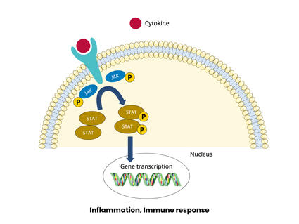
JAK Inhibitors Boost Immunotherapy in Clinical Trials
The combination shrank lymphoma and lung tumors in people and in mice.
Immune checkpoint inhibitor s are drugs that block an interaction between proteins on immune cells and cancer cells which, in turn, lowers the immune response to the cancer. Several immune checkpoint inhibitors have been approved for advanced lung cancer, including p embrolizumab (Keytruda) , a tezolizumab (Tecentriq) , c emiplimab (Libtayo) , d urvalumab (Imfinzi) , and n ivolumab (Opdivo) .
A key issue with immunotherapies is deciding which patients are most likely to benefit. There is some evidence that patients whose tumor cells have high levels of an immune checkpoint protein called PD-L1 may be more responsive to immune checkpoint inhibitors. Another marker for immunotherapy response is tumor mutational burden , or TMB, which refers to the amount of mutations in the DNA of the cancer cells. In some lung cancer trials, positive responses to immune checkpoint inhibitors have been linked with a high TMB. However, these markers cannot always predict a response and there is ongoing work to find better markers.
To learn more, see Immunotherapy to Treat Cancer .
Targeted Therapies
Targeted treatments identify and attack certain types of cancer cells with less harm to normal cells. In recent years, many targeted therapies have become available for advanced lung cancer and more are in development. Targeted treatments for lung cancer include the below.
Anaplastic lymphoma kinase (ALK) Inhibitors
ALK inhibitors target cancer-causing rearrangements in a protein called ALK. These drugs continue to be refined for the 5% of NSCLC patients who have an ALK gene alteration. Approved treatments include ceritinib (Zykadia) , alectinib (Alecensa) , brigatinib (Alunbrig) , and lorlatinib (Lorbrena) .
These ALK inhibitors are improvements from previous ones in their enhanced ability to cross the blood–brain barrier. This progress is critical because, in non-small cell lung cancer patients with ALK alterations, disease progression tends to occur in the brain. Based on clinical trial results, in 2024 the FDA approved alectinib as adjuvant therapy for people with ALK-positive NSCLC .
EGFR Inhibitors
Lung cancer trial of osimertinib draws praise—and some criticism.
The drug improved survival in a large clinical trial, but some question the trial’s design.
EGFR inhibitors block the activity of a protein called epidermal growth factor receptor (EGFR). Altered forms of EGFR are found at high levels in some lung cancers, causing them to grow rapidly. Osimertinib (Tagrisso) is the most effective and most widely used EGFR inhibitor. It is also used for adjuvant therapy after surgery for resectable NSCLC. Other drugs that target EGFR that are approved for treating NSCLC include afatinib (Gilotrif) , dacomitinib (Vizimpro) , erlotinib (Tarceva) , gefitinib (Iressa) . For people with Exon 20 mutations, amivantamab (Rybrevant) is an approved targeted therapy.
ROS1 Inhibitors
The ROS1 protein is involved in cell signaling and cell growth. A small percentage of people with NSCLC have rearranged forms of the ROS1 gene. Crizotinib (Xalkori) and entrectinib (Rozlytrek) are approved as treatments for patients with these alterations. In late 2023, the FDA approved repotrectinib (Augtyro) for advanced or metastatic NSCLC with ROS1 fusions as an initial treatment and as a second-line treatment in those who previously received a ROS1-targeted drug.
BRAF Inhibitors
The B-Raf protein is involved in sending signals in cells and cell growth. Certain changes in the B-Raf gene can increase the growth and spread of NSCLC cells.
The combination of the B-Raf-targeted drug dabrafenib (Tafinlar) and trametinib (Mekinist ), which targets a protein called MEK, has been approved as treatment for patients with NSCLC that has a specific mutation in the BRAF gene.
Encorafenib (Braftovi) combined with binimetinib (Mektovi) is approved for patients with metastatic NSCLC with a BRAF V600E mutation .
Other Inhibitors
Some NSCLCs have mutations in the genes NRTK-1 and NRTK-2 that can be treated with the targeted therapy larotrectinib (Vitrakvi). Those with certain mutations in the MET gene can be treated with tepotinib (Tepmetko) or capmatinib (Tabrecta) . And those with alterations in the RET gene are treated with selpercatinib (Retevmo) and pralsetinib (Gavreto) . A 2023 clinical trial showed that treatment with selpercatinib led to longer progression-free survival compared with people who received chemotherapy with or without pembrolizumab. Inhibitors of other targets that drive some lung cancers are being tested in clinical trials.
See a complete list of targeted therapies for lung cancer .
NCI-Supported Research Programs
Many NCI-funded researchers at the NIH campus, and across the United States and the world, are seeking ways to address lung cancer more effectively. Some research is basic, exploring questions as diverse as the biological underpinnings of cancer and the social factors that affect cancer risk. And some is more clinical, seeking to translate basic information into improved patient outcomes. The programs listed below are a small sampling of NCI’s research efforts in lung cancer.
- The Pragmatica-Lung Study is a randomized trial that will compare the combination of the targeted therapy ramucirumab (Cyramza) and the immunotherapy pembrolizumab (Keytruda) with standard chemotherapy in people with advanced NSCLC whose disease has progressed after previous treatment with immunotherapy and chemotherapy. In addition to looking at an important clinical question, the trial will serve as a model for future trials because it is designed to remove many of the barriers that prevent people from joining clinical trials.
- Begun in 2014, ALCHEMIST is a multicenter NCI trial for patients with early stage non-small cell lung cancer. It tests to see whether adding a targeted therapy after surgery, based on the genetics of a patient’s tumor, will improve survival.
- The Lung MAP trial is an ongoing multicenter trial for patients with advanced non-small cell lung cancer who have not responded to earlier treatment. Patients are assigned to specific targeted therapies based on their tumor’s genetic makeup.
- The Small Cell Lung Cancer Consortium was created to coordinate efforts and provide a network for investigators who focus on preclinical studies of small-cell lung cancer. The goal of the consortium is to accelerate progress on this disease through information exchange, data sharing and analysis, and face-to-face meetings.
- NCI funds eight lung cancer Specialized Programs of Research Excellence (Lung SPOREs) . These programs are designed to quickly move basic scientific findings into clinical settings. Each SPORE has multiple lung cancer projects underway.
Clinical Trials
NCI funds and oversees both early- and late-phase clinical trials to develop new treatments and improve patient care. Trials are available for both non-small cell lung cancer treatment and small cell lung cancer treatment .
Lung Cancer Research Results
The following are some of our latest news articles on lung cancer research:
- Lorlatinib Slows Growth of ALK-Positive Lung Cancers, May Prevent Brain Metastases
- Durvalumab Extends Lives of People with Early-Stage Small Cell Lung Cancer
- Alectinib Approved as an Adjuvant Treatment for Lung Cancer
- Repotrectinib Expands Treatment Options for Lung Cancers with ROS1 Fusions
- Tarlatamab Shows Promise for Some People with Small Cell Lung Cancer
- Selpercatinib Slows Progression of RET-Positive Lung, Medullary Thyroid Cancers
View the full list of Lung Cancer Research Results and Study Updates .

IMAGES
VIDEO
COMMENTS
case study on lung cancer - Free download as Word Doc (.doc / .docx), PDF File (.pdf), Text File (.txt) or view presentation slides online. The lungs are intricate organs made up of branching tubes and alveoli that facilitate gas exchange between inhaled air and blood. Within the alveoli, oxygen from the air passes into blood vessels while carbon dioxide moves out, carried by the blood to be ...
Case Study (Lung Cancer) - Free download as Word Doc (.doc / .docx), PDF File (.pdf), Text File (.txt) or read online for free. Lung cancer is classified into small cell lung cancer and non-small cell lung cancer, which is more common. NSCLC can be further divided into adenocarcinoma, squamous cell carcinoma, and large cell carcinoma. Smoking is the leading cause of lung cancer.
NUR 200 Lung Cancer Case Study (1) - Free download as Word Doc (.doc / .docx), PDF File (.pdf), Text File (.txt) or read online for free. Anniston, a 47-year-old smoker of 17 years, presents with hoarseness, difficulty swallowing, and coughing up rust-colored sputum. She is diagnosed with lung cancer. Diagnostic tests including chest x-ray, bronchoscopy, and CT scan are ordered.
Presentation of Case. Dr. Jonathan E. Eisen: A 47-year-old woman presented to this hospital early during the pandemic of coronavirus disease 2019 (Covid-19), the disease caused by severe acute ...
Dr. Mathew S. Lopes: A 65-year-old woman was transferred to this hospital because of chest pain. Six months before the current presentation, the patient presented to a hospital affiliated with ...
It's my pleasure to walk us through the first case, which is small cell lung cancer. This is a case with a 72-year-old woman who presents with shortness of breath, a productive cough, chest pain, some fatigue, anorexia, a recent 18-pound weight loss, and a history of hypertension. She is a schoolteacher and has a 45-pack-a-year smoking ...
Background. According to the Surveillance, Epidemiology, and End Results Program (SEER) registry based on 2007-2011 new cases, lung cancer (LC) is more frequently diagnosed among people aged 65-74 with only 1.6% of all cases occurring in patients younger than 45 years [].Most published data about LC in young populations are single-institutional retrospective analyses and few report on very ...
Case presentation. A 76-year-old man was referred for a lung mass in December 2018. He was a smoker (30 pack years with intermittent stops) and parking attendant for 30 years. There was no history of lung cancer in the immediate family of the patient. The patient was administered a dual bronchodilator for COPD.
According to 2004 WHO/International Association for the Study of Lung Cancer (IASLC) classification of lung and pleural tumors, CSCLC is defined as cancer tissues that mainly contain SCLC components with non-SCLC (NSCLC) histopathological types. The most common part of NSCLC is squamous cell carcinoma or large cell carcinoma (4, 5).
This document presents a case study of a 51-year-old man with small cell lung cancer. He received chemoradiotherapy followed by photodynamic therapy (PDT). After chemoradiotherapy, a mass remained in his left lung that was treated with PDT. Two years later, imaging and bronchoscopy showed a complete response with no recurrence of cancer. This represents a rare case where PDT led to a complete ...
Case presentation. A 76-year-old man was referred for a lung mass in December 2018. He was a smoker (30 pack years with intermittent stops) and parking attendant for 30 years. There was no history of lung cancer in the immediate family of the patient. The patient was administered a dual bronchodilator for COPD.
This is the first known report in the English literature to describe a case of metastatic non-small cell lung cancer that has been controlled for >11 years. ... phase III study of docetaxel plus platinum combinations versus vinorelbine plus cisplatin for advanced non-small-cell lung cancer: the TAX 326 study group. J Clin Oncol 2003;21:3016-24.
Here, we present the case of a 51-year-old man with limited-stage small cell lung cancer (LS-SCLC) who received concurrent chemoradiotherapy and photodynamic therapy (PDT). The patient was diagnosed as having LS-SCLC with an endobronchial mass in the left main bronchus. Following concurrent chemoradiotherapy, a mass remaining in the left lingular division was treated with PDT. Clinical and ...
Presentation of Case. Dr. Mathew S. Lopes: A 65-year-old woman was transferred to this hospital because of chest pain. Six months before the current presentation, the patient presented to a ...
In the presented study, we report a case of a 75-year-old female patient with advanced SCLC and an ECOG PS of 2 who responded well to combination immunochemotherapy with atezolizumab, etoposide, and carboplatin. ... Wu Y, Liu Y, Sun C, Immunotherapy as a treatment for small cell lung cancer: A case report and brief review: Transl Lung Cancer ...
This document provides background information on a case study about a male patient diagnosed with lung cancer. It discusses the patient's history, including his symptoms of shortness of breath, easy fatigability, and weight loss. It also covers the patient's family history, initial workup and diagnosis, and current hospital admission. The patient's health history, physical assessment ...
In this cross-sectional, case-control study, we examined various epidemiological factors and psychiatric comorbidities in primary and secondary lung cancer. Previous case-control studies on ...
The present study reported an extremely rare case of a 66-year-old male with non-small lung cell cancer in the left lobe and synchronous small cell lung cancer in the right lobe. The diagnosis of multiple primary lung cancer not only depends on biopsy pathology, but also requires molecular biology results. This is of great significance for the ...
Postsurgical clinical management for lung cancer is similar to that for all thoracic surgeries. Although surgery is reserved for clients with early-stage lung cancer, when it is indicated, chest tube management, pain control, mobilization, and venous thromboembolic (VTE) prophylaxis remain clinical priorities (Hoy et al., 2019).
Lung Ca Case study - Free download as PDF File (.pdf), Text File (.txt) or read online for free. Ms. Smith, a 45-year-old female smoker, presented with complaints of persistent cough, nausea, vomiting, poor appetite, weight loss, and pale skin. Objective findings included vital signs within normal limits, clear lungs, and concentrated urine.
Lung Cancer Case Study - Free download as Powerpoint Presentation (.ppt / .pptx), PDF File (.pdf), Text File (.txt) or view presentation slides online. A case Study regarding lung-cancer and the subtitle treatment for it giving an introduction at 1st then go through it step by step for best Evidence based practice
The NCI-sponsored National Lung Screening Trial (NLST) showed that low-dose CT scans can be used to screen for lung cancer in people with a history of heavy smoking. Using this screening can decrease their risk of dying from lung cancer. Now researchers are looking for ways to refine CT screening to better predict whether cancer is present.