The Power of Imaging: Why I am Passionate about Becoming a Sonographer
Many individuals who have been introduced to the world of diagnostic medical sonography have discovered the fascinating nature of this field and how the technology involved has evolved. As a result, their desire to pursue a career in this constantly evolving and exciting field has become more solidified. Discover the inspiration behind sonography and learn how to craft a compelling essay detailing your motivations for pursuing a career in this field. Our custom essay writing company offers tips and guidance on perfecting your essays, including, for instance, Why I Want to Become an Ultrasound Technician Essay.

Why I Want to be a Sonographer
My desire to become a sonographer stems from my belief that this role plays a crucial part in the healthcare system. With the use of ultrasound technology, sonographers capture images of the body that assist doctors in diagnosing various medical conditions. As a sonographer, I envision myself working directly with patients, providing them with the care and attention they need to make a positive impact on their healthcare outcomes. Furthermore, I view this career path as an opportunity to contribute to the overall well-being of the community.
Education and Training
To pursue a career as a sonographer, I understand that I must complete an accredited program in diagnostic medical sonography. Such a program comprises both didactic and clinical training, with the former providing me with a firm grasp of anatomy, physiology, and pathology, including the study of diseases. Through clinical training, I will gain hands-on experience in performing ultrasound scans and interacting with patients, thus preparing me for the certification exam and equipping me with the knowledge and skills needed to succeed in this field.
Job Opportunities
One of the advantages of pursuing a career as a sonographer is the diverse range of job opportunities available. Sonographers have the flexibility to work in various settings, including hospitals, clinics, and private practices. Moreover, the field offers specialized areas such as obstetrics, neurosonography, and vascular sonography, which provide additional career options. Additionally, the job outlook for sonographers is highly favorable, with the Bureau of Labor Statistics projecting a 23 percent increase in demand between 2016 and 2026. This makes it an appealing profession for individuals seeking job security and stability.
Advancements in Technology
The field of diagnostic medical sonography is constantly advancing, with continuous improvements in technology such as 3D and 4D imaging, making it an exciting time to consider becoming a sonographer. As a sonographer, I would have the opportunity to work with state-of-the-art equipment that can facilitate more precise and accurate diagnoses. Moreover, developments in portable ultrasound devices enable sonographers to reach patients in remote or inaccessible areas, thereby increasing access to care. As a result, I am convinced that the field of diagnostic medical sonography will continue to expand and evolve, presenting sonographers with outstanding opportunities for career growth and development.
Impact on Patients
The impact that sonographers can have on their patients is one of the most rewarding aspects of the field. By producing precise and timely images, sonographers can aid in early detection and diagnosis, ultimately improving patient outcomes and potentially saving lives. As someone who has personally experienced the anxiety and uncertainty that can accompany a medical diagnosis, I understand the value of having a healthcare provider who is empathetic and compassionate. In my role as a sonographer, I would have the opportunity to provide patients with the care and support they need during difficult and trying times, which is both humbling and fulfilling.
Personal and Professional Development
Finally, I am drawn to the field of sonography because it offers opportunities for personal and professional growth. As a lifelong learner, I appreciate that the field requires continuing education to stay up-to-date with developments in technology and best practices. Additionally, as I gain experience and expertise, I may have the opportunity to take on leadership roles or specialize in a particular area of sonography. I believe that this field will provide me with the opportunity to work in a challenging and rewarding role that allows me to make a difference in the lives of others while also fulfilling my own professional and personal goals.
As a pursuer of personal and professional development, I am deeply interested in the field of sonography. With this field, I have the potential to continuously learn and stay up-to-date with new technologies and techniques. As my career progresses, I can take on leadership roles or specialize in certain areas of sonography. I’m confident that this field will provide a fulfilling job that both helps others and caters to my individual goals.
Tips on Writing Essay “Why I Want to be a Sonographer”
To write a convincing why do you want to be a sonographer essay, it’s crucial to emphasize your reasons for choosing this profession. To make your essay stand out, focus on what excites you about the field and explain how it aligns with your goals and interests.
Also, don’t forget to mention any relevant experience or skills that make you a strong candidate. Demonstrating enthusiasm and passion for the field is vital to show your commitment to the profession. Ensure that your essay is well-organized, structured, descriptive, and engaging. To write an opinion essay on the topic, start with a clear and concise thesis statement, use specific examples and experiences to support your argument, address potential counterarguments, and use clear language that is easy for readers to understand.
Finally, conclude with a strong statement that summarizes your argument and leaves a lasting impression on the reader.
Related posts:
- What Does Rain Symbolize in Literature
- Healing with Heart: Essay About My Plans to Becoming a Doctor
- Why I Want to Become a Counselor, Essay Sample
- Democracy in Question: Should The Electoral College be Abolished Essay
Improve your writing with our guides

Youth Culture Essay Prompt and Discussion

Why Should College Athletes Be Paid, Essay Sample

Reasons Why Minimum Wage Should Be Raised Essay: Benefits for Workers, Society, and The Economy
Get 15% off your first order with edusson.
Connect with a professional writer within minutes by placing your first order. No matter the subject, difficulty, academic level or document type, our writers have the skills to complete it.
100% privacy. No spam ever.

Head Start Your Radiology Residency [Online] ↗️
- Radiology Thesis – More than 400 Research Topics (2022)!
Username or Email Address
Remember Me

Introduction
A thesis or dissertation, as some people would like to call it, is an integral part of the Radiology curriculum, be it MD, DNB, or DMRD. We have tried to aggregate radiology thesis topics from various sources for reference.
Not everyone is interested in research, and writing a Radiology thesis can be daunting. But there is no escape from preparing, so it is better that you accept this bitter truth and start working on it instead of cribbing about it (like other things in life. #PhilosophyGyan!)
Start working on your thesis as early as possible and finish your thesis well before your exams, so you do not have that stress at the back of your mind. Also, your thesis may need multiple revisions, so be prepared and allocate time accordingly.
Tips for Choosing Radiology Thesis and Research Topics
Keep it simple silly (kiss).
Retrospective > Prospective
Retrospective studies are better than prospective ones, as you already have the data you need when choosing to do a retrospective study. Prospective studies are better quality, but as a resident, you may not have time (, energy and enthusiasm) to complete these.
Choose a simple topic that answers a single/few questions
Original research is challenging, especially if you do not have prior experience. I would suggest you choose a topic that answers a single or few questions. Most topics that I have listed are along those lines. Alternatively, you can choose a broad topic such as “Role of MRI in evaluation of perianal fistulas.”
You can choose a novel topic if you are genuinely interested in research AND have a good mentor who will guide you. Once you have done that, make sure that you publish your study once you are done with it.
Get it done ASAP.
In most cases, it makes sense to stick to a thesis topic that will not take much time. That does not mean you should ignore your thesis and ‘Ctrl C + Ctrl V’ from a friend from another university. Thesis writing is your first step toward research methodology so do it as sincerely as possible. Do not procrastinate in preparing the thesis. As soon as you have been allotted a guide, start researching topics and writing a review of the literature.
At the same time, do not invest a lot of time in writing/collecting data for your thesis. You should not be busy finishing your thesis a few months before the exam. Some people could not appear for the exam because they could not submit their thesis in time. So DO NOT TAKE thesis lightly.
Do NOT Copy-Paste
Reiterating once again, do not simply choose someone else’s thesis topic. Find out what are kind of cases that your Hospital caters to. It is better to do a good thesis on a common topic than a crappy one on a rare one.
Books to help you write a Radiology Thesis
Event country/university has a different format for thesis; hence these book recommendations may not work for everyone.

- Amazon Kindle Edition
- Gupta, Piyush (Author)
- English (Publication Language)
- 206 Pages - 10/12/2020 (Publication Date) - Jaypee Brothers Medical Publishers (P) Ltd. (Publisher)
In A Hurry? Download a PDF list of Radiology Research Topics!
Sign up below to get this PDF directly to your email address.
100% Privacy Guaranteed. Your information will not be shared. Unsubscribe anytime with a single click.
List of Radiology Research /Thesis / Dissertation Topics
- State of the art of MRI in the diagnosis of hepatic focal lesions
- Multimodality imaging evaluation of sacroiliitis in newly diagnosed patients of spondyloarthropathy
- Multidetector computed tomography in oesophageal varices
- Role of positron emission tomography with computed tomography in the diagnosis of cancer Thyroid
- Evaluation of focal breast lesions using ultrasound elastography
- Role of MRI diffusion tensor imaging in the assessment of traumatic spinal cord injuries
- Sonographic imaging in male infertility
- Comparison of color Doppler and digital subtraction angiography in occlusive arterial disease in patients with lower limb ischemia
- The role of CT urography in Haematuria
- Role of functional magnetic resonance imaging in making brain tumor surgery safer
- Prediction of pre-eclampsia and fetal growth restriction by uterine artery Doppler
- Role of grayscale and color Doppler ultrasonography in the evaluation of neonatal cholestasis
- Validity of MRI in the diagnosis of congenital anorectal anomalies
- Role of sonography in assessment of clubfoot
- Role of diffusion MRI in preoperative evaluation of brain neoplasms
- Imaging of upper airways for pre-anaesthetic evaluation purposes and for laryngeal afflictions.
- A study of multivessel (arterial and venous) Doppler velocimetry in intrauterine growth restriction
- Multiparametric 3tesla MRI of suspected prostatic malignancy.
- Role of Sonography in Characterization of Thyroid Nodules for differentiating benign from
- Role of advances magnetic resonance imaging sequences in multiple sclerosis
- Role of multidetector computed tomography in evaluation of jaw lesions
- Role of Ultrasound and MR Imaging in the Evaluation of Musculotendinous Pathologies of Shoulder Joint
- Role of perfusion computed tomography in the evaluation of cerebral blood flow, blood volume and vascular permeability of cerebral neoplasms
- MRI flow quantification in the assessment of the commonest csf flow abnormalities
- Role of diffusion-weighted MRI in evaluation of prostate lesions and its histopathological correlation
- CT enterography in evaluation of small bowel disorders
- Comparison of perfusion magnetic resonance imaging (PMRI), magnetic resonance spectroscopy (MRS) in and positron emission tomography-computed tomography (PET/CT) in post radiotherapy treated gliomas to detect recurrence
- Role of multidetector computed tomography in evaluation of paediatric retroperitoneal masses
- Role of Multidetector computed tomography in neck lesions
- Estimation of standard liver volume in Indian population
- Role of MRI in evaluation of spinal trauma
- Role of modified sonohysterography in female factor infertility: a pilot study.
- The role of pet-CT in the evaluation of hepatic tumors
- Role of 3D magnetic resonance imaging tractography in assessment of white matter tracts compromise in supratentorial tumors
- Role of dual phase multidetector computed tomography in gallbladder lesions
- Role of multidetector computed tomography in assessing anatomical variants of nasal cavity and paranasal sinuses in patients of chronic rhinosinusitis.
- magnetic resonance spectroscopy in multiple sclerosis
- Evaluation of thyroid nodules by ultrasound elastography using acoustic radiation force impulse (ARFI) imaging
- Role of Magnetic Resonance Imaging in Intractable Epilepsy
- Evaluation of suspected and known coronary artery disease by 128 slice multidetector CT.
- Role of regional diffusion tensor imaging in the evaluation of intracranial gliomas and its histopathological correlation
- Role of chest sonography in diagnosing pneumothorax
- Role of CT virtual cystoscopy in diagnosis of urinary bladder neoplasia
- Role of MRI in assessment of valvular heart diseases
- High resolution computed tomography of temporal bone in unsafe chronic suppurative otitis media
- Multidetector CT urography in the evaluation of hematuria
- Contrast-induced nephropathy in diagnostic imaging investigations with intravenous iodinated contrast media
- Comparison of dynamic susceptibility contrast-enhanced perfusion magnetic resonance imaging and single photon emission computed tomography in patients with little’s disease
- Role of Multidetector Computed Tomography in Bowel Lesions.
- Role of diagnostic imaging modalities in evaluation of post liver transplantation recipient complications.
- Role of multislice CT scan and barium swallow in the estimation of oesophageal tumour length
- Malignant Lesions-A Prospective Study.
- Value of ultrasonography in assessment of acute abdominal diseases in pediatric age group
- Role of three dimensional multidetector CT hysterosalpingography in female factor infertility
- Comparative evaluation of multi-detector computed tomography (MDCT) virtual tracheo-bronchoscopy and fiberoptic tracheo-bronchoscopy in airway diseases
- Role of Multidetector CT in the evaluation of small bowel obstruction
- Sonographic evaluation in adhesive capsulitis of shoulder
- Utility of MR Urography Versus Conventional Techniques in Obstructive Uropathy
- MRI of the postoperative knee
- Role of 64 slice-multi detector computed tomography in diagnosis of bowel and mesenteric injury in blunt abdominal trauma.
- Sonoelastography and triphasic computed tomography in the evaluation of focal liver lesions
- Evaluation of Role of Transperineal Ultrasound and Magnetic Resonance Imaging in Urinary Stress incontinence in Women
- Multidetector computed tomographic features of abdominal hernias
- Evaluation of lesions of major salivary glands using ultrasound elastography
- Transvaginal ultrasound and magnetic resonance imaging in female urinary incontinence
- MDCT colonography and double-contrast barium enema in evaluation of colonic lesions
- Role of MRI in diagnosis and staging of urinary bladder carcinoma
- Spectrum of imaging findings in children with febrile neutropenia.
- Spectrum of radiographic appearances in children with chest tuberculosis.
- Role of computerized tomography in evaluation of mediastinal masses in pediatric
- Diagnosing renal artery stenosis: Comparison of multimodality imaging in diabetic patients
- Role of multidetector CT virtual hysteroscopy in the detection of the uterine & tubal causes of female infertility
- Role of multislice computed tomography in evaluation of crohn’s disease
- CT quantification of parenchymal and airway parameters on 64 slice MDCT in patients of chronic obstructive pulmonary disease
- Comparative evaluation of MDCT and 3t MRI in radiographically detected jaw lesions.
- Evaluation of diagnostic accuracy of ultrasonography, colour Doppler sonography and low dose computed tomography in acute appendicitis
- Ultrasonography , magnetic resonance cholangio-pancreatography (MRCP) in assessment of pediatric biliary lesions
- Multidetector computed tomography in hepatobiliary lesions.
- Evaluation of peripheral nerve lesions with high resolution ultrasonography and colour Doppler
- Multidetector computed tomography in pancreatic lesions
- Multidetector Computed Tomography in Paediatric abdominal masses.
- Evaluation of focal liver lesions by colour Doppler and MDCT perfusion imaging
- Sonographic evaluation of clubfoot correction during Ponseti treatment
- Role of multidetector CT in characterization of renal masses
- Study to assess the role of Doppler ultrasound in evaluation of arteriovenous (av) hemodialysis fistula and the complications of hemodialysis vasular access
- Comparative study of multiphasic contrast-enhanced CT and contrast-enhanced MRI in the evaluation of hepatic mass lesions
- Sonographic spectrum of rheumatoid arthritis
- Diagnosis & staging of liver fibrosis by ultrasound elastography in patients with chronic liver diseases
- Role of multidetector computed tomography in assessment of jaw lesions.
- Role of high-resolution ultrasonography in the differentiation of benign and malignant thyroid lesions
- Radiological evaluation of aortic aneurysms in patients selected for endovascular repair
- Role of conventional MRI, and diffusion tensor imaging tractography in evaluation of congenital brain malformations
- To evaluate the status of coronary arteries in patients with non-valvular atrial fibrillation using 256 multirow detector CT scan
- A comparative study of ultrasonography and CT – arthrography in diagnosis of chronic ligamentous and meniscal injuries of knee
- Multi detector computed tomography evaluation in chronic obstructive pulmonary disease and correlation with severity of disease
- Diffusion weighted and dynamic contrast enhanced magnetic resonance imaging in chemoradiotherapeutic response evaluation in cervical cancer.
- High resolution sonography in the evaluation of non-traumatic painful wrist
- The role of trans-vaginal ultrasound versus magnetic resonance imaging in diagnosis & evaluation of cancer cervix
- Role of multidetector row computed tomography in assessment of maxillofacial trauma
- Imaging of vascular complication after liver transplantation.
- Role of magnetic resonance perfusion weighted imaging & spectroscopy for grading of glioma by correlating perfusion parameter of the lesion with the final histopathological grade
- Magnetic resonance evaluation of abdominal tuberculosis.
- Diagnostic usefulness of low dose spiral HRCT in diffuse lung diseases
- Role of dynamic contrast enhanced and diffusion weighted magnetic resonance imaging in evaluation of endometrial lesions
- Contrast enhanced digital mammography anddigital breast tomosynthesis in early diagnosis of breast lesion
- Evaluation of Portal Hypertension with Colour Doppler flow imaging and magnetic resonance imaging
- Evaluation of musculoskeletal lesions by magnetic resonance imaging
- Role of diffusion magnetic resonance imaging in assessment of neoplastic and inflammatory brain lesions
- Radiological spectrum of chest diseases in HIV infected children High resolution ultrasonography in neck masses in children
- with surgical findings
- Sonographic evaluation of peripheral nerves in type 2 diabetes mellitus.
- Role of perfusion computed tomography in the evaluation of neck masses and correlation
- Role of ultrasonography in the diagnosis of knee joint lesions
- Role of ultrasonography in evaluation of various causes of pelvic pain in first trimester of pregnancy.
- Role of Magnetic Resonance Angiography in the Evaluation of Diseases of Aorta and its Branches
- MDCT fistulography in evaluation of fistula in Ano
- Role of multislice CT in diagnosis of small intestine tumors
- Role of high resolution CT in differentiation between benign and malignant pulmonary nodules in children
- A study of multidetector computed tomography urography in urinary tract abnormalities
- Role of high resolution sonography in assessment of ulnar nerve in patients with leprosy.
- Pre-operative radiological evaluation of locally aggressive and malignant musculoskeletal tumours by computed tomography and magnetic resonance imaging.
- The role of ultrasound & MRI in acute pelvic inflammatory disease
- Ultrasonography compared to computed tomographic arthrography in the evaluation of shoulder pain
- Role of Multidetector Computed Tomography in patients with blunt abdominal trauma.
- The Role of Extended field-of-view Sonography and compound imaging in Evaluation of Breast Lesions
- Evaluation of focal pancreatic lesions by Multidetector CT and perfusion CT
- Evaluation of breast masses on sono-mammography and colour Doppler imaging
- Role of CT virtual laryngoscopy in evaluation of laryngeal masses
- Triple phase multi detector computed tomography in hepatic masses
- Role of transvaginal ultrasound in diagnosis and treatment of female infertility
- Role of ultrasound and color Doppler imaging in assessment of acute abdomen due to female genetal causes
- High resolution ultrasonography and color Doppler ultrasonography in scrotal lesion
- Evaluation of diagnostic accuracy of ultrasonography with colour Doppler vs low dose computed tomography in salivary gland disease
- Role of multidetector CT in diagnosis of salivary gland lesions
- Comparison of diagnostic efficacy of ultrasonography and magnetic resonance cholangiopancreatography in obstructive jaundice: A prospective study
- Evaluation of varicose veins-comparative assessment of low dose CT venogram with sonography: pilot study
- Role of mammotome in breast lesions
- The role of interventional imaging procedures in the treatment of selected gynecological disorders
- Role of transcranial ultrasound in diagnosis of neonatal brain insults
- Role of multidetector CT virtual laryngoscopy in evaluation of laryngeal mass lesions
- Evaluation of adnexal masses on sonomorphology and color Doppler imaginig
- Role of radiological imaging in diagnosis of endometrial carcinoma
- Comprehensive imaging of renal masses by magnetic resonance imaging
- The role of 3D & 4D ultrasonography in abnormalities of fetal abdomen
- Diffusion weighted magnetic resonance imaging in diagnosis and characterization of brain tumors in correlation with conventional MRI
- Role of diffusion weighted MRI imaging in evaluation of cancer prostate
- Role of multidetector CT in diagnosis of urinary bladder cancer
- Role of multidetector computed tomography in the evaluation of paediatric retroperitoneal masses.
- Comparative evaluation of gastric lesions by double contrast barium upper G.I. and multi detector computed tomography
- Evaluation of hepatic fibrosis in chronic liver disease using ultrasound elastography
- Role of MRI in assessment of hydrocephalus in pediatric patients
- The role of sonoelastography in characterization of breast lesions
- The influence of volumetric tumor doubling time on survival of patients with intracranial tumours
- Role of perfusion computed tomography in characterization of colonic lesions
- Role of proton MRI spectroscopy in the evaluation of temporal lobe epilepsy
- Role of Doppler ultrasound and multidetector CT angiography in evaluation of peripheral arterial diseases.
- Role of multidetector computed tomography in paranasal sinus pathologies
- Role of virtual endoscopy using MDCT in detection & evaluation of gastric pathologies
- High resolution 3 Tesla MRI in the evaluation of ankle and hindfoot pain.
- Transperineal ultrasonography in infants with anorectal malformation
- CT portography using MDCT versus color Doppler in detection of varices in cirrhotic patients
- Role of CT urography in the evaluation of a dilated ureter
- Characterization of pulmonary nodules by dynamic contrast-enhanced multidetector CT
- Comprehensive imaging of acute ischemic stroke on multidetector CT
- The role of fetal MRI in the diagnosis of intrauterine neurological congenital anomalies
- Role of Multidetector computed tomography in pediatric chest masses
- Multimodality imaging in the evaluation of palpable & non-palpable breast lesion.
- Sonographic Assessment Of Fetal Nasal Bone Length At 11-28 Gestational Weeks And Its Correlation With Fetal Outcome.
- Role Of Sonoelastography And Contrast-Enhanced Computed Tomography In Evaluation Of Lymph Node Metastasis In Head And Neck Cancers
- Role Of Renal Doppler And Shear Wave Elastography In Diabetic Nephropathy
- Evaluation Of Relationship Between Various Grades Of Fatty Liver And Shear Wave Elastography Values
- Evaluation and characterization of pelvic masses of gynecological origin by USG, color Doppler and MRI in females of reproductive age group
- Radiological evaluation of small bowel diseases using computed tomographic enterography
- Role of coronary CT angiography in patients of coronary artery disease
- Role of multimodality imaging in the evaluation of pediatric neck masses
- Role of CT in the evaluation of craniocerebral trauma
- Role of magnetic resonance imaging (MRI) in the evaluation of spinal dysraphism
- Comparative evaluation of triple phase CT and dynamic contrast-enhanced MRI in patients with liver cirrhosis
- Evaluation of the relationship between carotid intima-media thickness and coronary artery disease in patients evaluated by coronary angiography for suspected CAD
- Assessment of hepatic fat content in fatty liver disease by unenhanced computed tomography
- Correlation of vertebral marrow fat on spectroscopy and diffusion-weighted MRI imaging with bone mineral density in postmenopausal women.
- Comparative evaluation of CT coronary angiography with conventional catheter coronary angiography
- Ultrasound evaluation of kidney length & descending colon diameter in normal and intrauterine growth-restricted fetuses
- A prospective study of hepatic vein waveform and splenoportal index in liver cirrhosis: correlation with child Pugh’s classification and presence of esophageal varices.
- CT angiography to evaluate coronary artery by-pass graft patency in symptomatic patient’s functional assessment of myocardium by cardiac MRI in patients with myocardial infarction
- MRI evaluation of HIV positive patients with central nervous system manifestations
- MDCT evaluation of mediastinal and hilar masses
- Evaluation of rotator cuff & labro-ligamentous complex lesions by MRI & MRI arthrography of shoulder joint
- Role of imaging in the evaluation of soft tissue vascular malformation
- Role of MRI and ultrasonography in the evaluation of multifidus muscle pathology in chronic low back pain patients
- Role of ultrasound elastography in the differential diagnosis of breast lesions
- Role of magnetic resonance cholangiopancreatography in evaluating dilated common bile duct in patients with symptomatic gallstone disease.
- Comparative study of CT urography & hybrid CT urography in patients with haematuria.
- Role of MRI in the evaluation of anorectal malformations
- Comparison of ultrasound-Doppler and magnetic resonance imaging findings in rheumatoid arthritis of hand and wrist
- Role of Doppler sonography in the evaluation of renal artery stenosis in hypertensive patients undergoing coronary angiography for coronary artery disease.
- Comparison of radiography, computed tomography and magnetic resonance imaging in the detection of sacroiliitis in ankylosing spondylitis.
- Mr evaluation of painful hip
- Role of MRI imaging in pretherapeutic assessment of oral and oropharyngeal malignancy
- Evaluation of diffuse lung diseases by high resolution computed tomography of the chest
- Mr evaluation of brain parenchyma in patients with craniosynostosis.
- Diagnostic and prognostic value of cardiovascular magnetic resonance imaging in dilated cardiomyopathy
- Role of multiparametric magnetic resonance imaging in the detection of early carcinoma prostate
- Role of magnetic resonance imaging in white matter diseases
- Role of sonoelastography in assessing the response to neoadjuvant chemotherapy in patients with locally advanced breast cancer.
- Role of ultrasonography in the evaluation of carotid and femoral intima-media thickness in predialysis patients with chronic kidney disease
- Role of H1 MRI spectroscopy in focal bone lesions of peripheral skeleton choline detection by MRI spectroscopy in breast cancer and its correlation with biomarkers and histological grade.
- Ultrasound and MRI evaluation of axillary lymph node status in breast cancer.
- Role of sonography and magnetic resonance imaging in evaluating chronic lateral epicondylitis.
- Comparative of sonography including Doppler and sonoelastography in cervical lymphadenopathy.
- Evaluation of Umbilical Coiling Index as Predictor of Pregnancy Outcome.
- Computerized Tomographic Evaluation of Azygoesophageal Recess in Adults.
- Lumbar Facet Arthropathy in Low Backache.
- “Urethral Injuries After Pelvic Trauma: Evaluation with Uretrography
- Role Of Ct In Diagnosis Of Inflammatory Renal Diseases
- Role Of Ct Virtual Laryngoscopy In Evaluation Of Laryngeal Masses
- “Ct Portography Using Mdct Versus Color Doppler In Detection Of Varices In
- Cirrhotic Patients”
- Role Of Multidetector Ct In Characterization Of Renal Masses
- Role Of Ct Virtual Cystoscopy In Diagnosis Of Urinary Bladder Neoplasia
- Role Of Multislice Ct In Diagnosis Of Small Intestine Tumors
- “Mri Flow Quantification In The Assessment Of The Commonest CSF Flow Abnormalities”
- “The Role Of Fetal Mri In Diagnosis Of Intrauterine Neurological CongenitalAnomalies”
- Role Of Transcranial Ultrasound In Diagnosis Of Neonatal Brain Insults
- “The Role Of Interventional Imaging Procedures In The Treatment Of Selected Gynecological Disorders”
- Role Of Radiological Imaging In Diagnosis Of Endometrial Carcinoma
- “Role Of High-Resolution Ct In Differentiation Between Benign And Malignant Pulmonary Nodules In Children”
- Role Of Ultrasonography In The Diagnosis Of Knee Joint Lesions
- “Role Of Diagnostic Imaging Modalities In Evaluation Of Post Liver Transplantation Recipient Complications”
- “Diffusion-Weighted Magnetic Resonance Imaging In Diagnosis And
- Characterization Of Brain Tumors In Correlation With Conventional Mri”
- The Role Of PET-CT In The Evaluation Of Hepatic Tumors
- “Role Of Computerized Tomography In Evaluation Of Mediastinal Masses In Pediatric patients”
- “Trans Vaginal Ultrasound And Magnetic Resonance Imaging In Female Urinary Incontinence”
- Role Of Multidetector Ct In Diagnosis Of Urinary Bladder Cancer
- “Role Of Transvaginal Ultrasound In Diagnosis And Treatment Of Female Infertility”
- Role Of Diffusion-Weighted Mri Imaging In Evaluation Of Cancer Prostate
- “Role Of Positron Emission Tomography With Computed Tomography In Diagnosis Of Cancer Thyroid”
- The Role Of CT Urography In Case Of Haematuria
- “Value Of Ultrasonography In Assessment Of Acute Abdominal Diseases In Pediatric Age Group”
- “Role Of Functional Magnetic Resonance Imaging In Making Brain Tumor Surgery Safer”
- The Role Of Sonoelastography In Characterization Of Breast Lesions
- “Ultrasonography, Magnetic Resonance Cholangiopancreatography (MRCP) In Assessment Of Pediatric Biliary Lesions”
- “Role Of Ultrasound And Color Doppler Imaging In Assessment Of Acute Abdomen Due To Female Genital Causes”
- “Role Of Multidetector Ct Virtual Laryngoscopy In Evaluation Of Laryngeal Mass Lesions”
- MRI Of The Postoperative Knee
- Role Of Mri In Assessment Of Valvular Heart Diseases
- The Role Of 3D & 4D Ultrasonography In Abnormalities Of Fetal Abdomen
- State Of The Art Of Mri In Diagnosis Of Hepatic Focal Lesions
- Role Of Multidetector Ct In Diagnosis Of Salivary Gland Lesions
- “Role Of Virtual Endoscopy Using Mdct In Detection & Evaluation Of Gastric Pathologies”
- The Role Of Ultrasound & Mri In Acute Pelvic Inflammatory Disease
- “Diagnosis & Staging Of Liver Fibrosis By Ultraso Und Elastography In
- Patients With Chronic Liver Diseases”
- Role Of Mri In Evaluation Of Spinal Trauma
- Validity Of Mri In Diagnosis Of Congenital Anorectal Anomalies
- Imaging Of Vascular Complication After Liver Transplantation
- “Contrast-Enhanced Digital Mammography And Digital Breast Tomosynthesis In Early Diagnosis Of Breast Lesion”
- Role Of Mammotome In Breast Lesions
- “Role Of MRI Diffusion Tensor Imaging (DTI) In Assessment Of Traumatic Spinal Cord Injuries”
- “Prediction Of Pre-eclampsia And Fetal Growth Restriction By Uterine Artery Doppler”
- “Role Of Multidetector Row Computed Tomography In Assessment Of Maxillofacial Trauma”
- “Role Of Diffusion Magnetic Resonance Imaging In Assessment Of Neoplastic And Inflammatory Brain Lesions”
- Role Of Diffusion Mri In Preoperative Evaluation Of Brain Neoplasms
- “Role Of Multidetector Ct Virtual Hysteroscopy In The Detection Of The
- Uterine & Tubal Causes Of Female Infertility”
- Role Of Advances Magnetic Resonance Imaging Sequences In Multiple Sclerosis Magnetic Resonance Spectroscopy In Multiple Sclerosis
- “Role Of Conventional Mri, And Diffusion Tensor Imaging Tractography In Evaluation Of Congenital Brain Malformations”
- Role Of MRI In Evaluation Of Spinal Trauma
- Diagnostic Role Of Diffusion-weighted MR Imaging In Neck Masses
- “The Role Of Transvaginal Ultrasound Versus Magnetic Resonance Imaging In Diagnosis & Evaluation Of Cancer Cervix”
- “Role Of 3d Magnetic Resonance Imaging Tractography In Assessment Of White Matter Tracts Compromise In Supra Tentorial Tumors”
- Role Of Proton MR Spectroscopy In The Evaluation Of Temporal Lobe Epilepsy
- Role Of Multislice Computed Tomography In Evaluation Of Crohn’s Disease
- Role Of MRI In Assessment Of Hydrocephalus In Pediatric Patients
- The Role Of MRI In Diagnosis And Staging Of Urinary Bladder Carcinoma
- USG and MRI correlation of congenital CNS anomalies
- HRCT in interstitial lung disease
- X-Ray, CT and MRI correlation of bone tumors
- “Study on the diagnostic and prognostic utility of X-Rays for cases of pulmonary tuberculosis under RNTCP”
- “Role of magnetic resonance imaging in the characterization of female adnexal pathology”
- “CT angiography of carotid atherosclerosis and NECT brain in cerebral ischemia, a correlative analysis”
- Role of CT scan in the evaluation of paranasal sinus pathology
- USG and MRI correlation on shoulder joint pathology
- “Radiological evaluation of a patient presenting with extrapulmonary tuberculosis”
- CT and MRI correlation in focal liver lesions”
- Comparison of MDCT virtual cystoscopy with conventional cystoscopy in bladder tumors”
- “Bleeding vessels in life-threatening hemoptysis: Comparison of 64 detector row CT angiography with conventional angiography prior to endovascular management”
- “Role of transarterial chemoembolization in unresectable hepatocellular carcinoma”
- “Comparison of color flow duplex study with digital subtraction angiography in the evaluation of peripheral vascular disease”
- “A Study to assess the efficacy of magnetization transfer ratio in differentiating tuberculoma from neurocysticercosis”
- “MR evaluation of uterine mass lesions in correlation with transabdominal, transvaginal ultrasound using HPE as a gold standard”
- “The Role of power Doppler imaging with trans rectal ultrasonogram guided prostate biopsy in the detection of prostate cancer”
- “Lower limb arteries assessed with doppler angiography – A prospective comparative study with multidetector CT angiography”
- “Comparison of sildenafil with papaverine in penile doppler by assessing hemodynamic changes”
- “Evaluation of efficacy of sonosalphingogram for assessing tubal patency in infertile patients with hysterosalpingogram as the gold standard”
- Role of CT enteroclysis in the evaluation of small bowel diseases
- “MRI colonography versus conventional colonoscopy in the detection of colonic polyposis”
- “Magnetic Resonance Imaging of anteroposterior diameter of the midbrain – differentiation of progressive supranuclear palsy from Parkinson disease”
- “MRI Evaluation of anterior cruciate ligament tears with arthroscopic correlation”
- “The Clinicoradiological profile of cerebral venous sinus thrombosis with prognostic evaluation using MR sequences”
- “Role of MRI in the evaluation of pelvic floor integrity in stress incontinent patients” “Doppler ultrasound evaluation of hepatic venous waveform in portal hypertension before and after propranolol”
- “Role of transrectal sonography with colour doppler and MRI in evaluation of prostatic lesions with TRUS guided biopsy correlation”
- “Ultrasonographic evaluation of painful shoulders and correlation of rotator cuff pathologies and clinical examination”
- “Colour Doppler Evaluation of Common Adult Hepatic tumors More Than 2 Cm with HPE and CECT Correlation”
- “Clinical Relevance of MR Urethrography in Obliterative Posterior Urethral Stricture”
- “Prediction of Adverse Perinatal Outcome in Growth Restricted Fetuses with Antenatal Doppler Study”
- Radiological evaluation of spinal dysraphism using CT and MRI
- “Evaluation of temporal bone in cholesteatoma patients by high resolution computed tomography”
- “Radiological evaluation of primary brain tumours using computed tomography and magnetic resonance imaging”
- “Three dimensional colour doppler sonographic assessment of changes in volume and vascularity of fibroids – before and after uterine artery embolization”
- “In phase opposed phase imaging of bone marrow differentiating neoplastic lesions”
- “Role of dynamic MRI in replacing the isotope renogram in the functional evaluation of PUJ obstruction”
- Characterization of adrenal masses with contrast-enhanced CT – washout study
- A study on accuracy of magnetic resonance cholangiopancreatography
- “Evaluation of median nerve in carpal tunnel syndrome by high-frequency ultrasound & color doppler in comparison with nerve conduction studies”
- “Correlation of Agatston score in patients with obstructive and nonobstructive coronary artery disease following STEMI”
- “Doppler ultrasound assessment of tumor vascularity in locally advanced breast cancer at diagnosis and following primary systemic chemotherapy.”
- “Validation of two-dimensional perineal ultrasound and dynamic magnetic resonance imaging in pelvic floor dysfunction.”
- “Role of MR urethrography compared to conventional urethrography in the surgical management of obliterative urethral stricture.”
Search Diagnostic Imaging Research Topics
You can also search research-related resources and direct download PDFs for radiology articles on our custom radiology search engine .

Free Resources for Preparing Radiology Thesis
- Radiology thesis topics- Benha University – Free to download thesis
- Radiology thesis topics – Faculty of Medical Science Delhi
- Radiology thesis topics – IPGMER
- Fetal Radiology thesis Protocols
- Radiology thesis and dissertation topics
- Radiographics
Proofreading Your Thesis:
Make sure you use Grammarly to correct your spelling , grammar , and plagiarism for your thesis. Grammarly has affordable paid subscriptions, windows/macOS apps, and FREE browser extensions. It is an excellent tool to avoid inadvertent spelling mistakes in your research projects. It has an extensive built-in vocabulary, but you should make an account and add your own medical glossary to it.

Guidelines for Writing a Radiology Thesis:
These are general guidelines and not about radiology specifically. You can share these with colleagues from other departments as well. Special thanks to Dr. Sanjay Yadav sir for these. This section is best seen on a desktop. Here are a couple of handy presentations to start writing a thesis:
Read the general guidelines for writing a thesis (the page will take some time to load- more than 70 pages!
A format for thesis protocol with a sample patient information sheet, sample patient consent form, sample application letter for thesis, and sample certificate.
Resources and References:
- Guidelines for thesis writing.
- Format for thesis protocol
- Thesis protocol writing guidelines DNB
- Informed consent form for Research studies from AIIMS
- Radiology Informed consent forms in local Indian languages.
- Sample Informed Consent form for Research in Hindi
- Guide to write a thesis by Dr. P R Sharma
- Guidelines for thesis writing by Dr. Pulin Gupta.
- Preparing MD/DNB thesis by A Indrayan
- Another good thesis reference protocol
Hopefully, this post will make the tedious task of writing a Radiology thesis a little bit easier for you. Best of luck with writing your thesis and your residency too!
More guides for residents :
- Guide for the MD/DMRD/DNB radiology exam!
- Guide for First-Year Radiology Residents
- FRCR Exam: THE Most Comprehensive Guide (2022)!
- Radiology Practical Exams Questions compilation for MD/DNB/DMRD !
- Radiology Exam Resources (Oral Recalls, Instruments, etc )!
- Tips and Tricks for DNB/MD Radiology Practical Exam
- FRCR 2B exam- Tips and Tricks !
FRCR exam preparation – An alternative take!
- Why did I take up Radiology?
- Radiology Conferences – A comprehensive guide!
- ECR (European Congress Of Radiology)
- European Diploma in Radiology (EDiR) – The Complete Guide!
- Radiology NEET PG guide – How to select THE best college for post-graduation in Radiology (includes personal insights)!
Interventional Radiology – All Your Questions Answered!
- What It Means To Be A Radiologist: A Guide For Medical Students!
- Radiology Mentors for Medical Students (Post NEET-PG)
- MD vs DNB Radiology: Which Path is Right for Your Career?
- DNB Radiology OSCE – Tips and Tricks
More radiology resources here: Radiology resources This page will be updated regularly. Kindly leave your feedback in the comments or send us a message here . Also, you can comment below regarding your department’s thesis topics.
Note: All topics have been compiled from available online resources. If anyone has an issue with any radiology thesis topics displayed here, you can message us here , and we can delete them. These are only sample guidelines. Thesis guidelines differ from institution to institution.
Image source: Thesis complete! (2018). Flickr. Retrieved 12 August 2018, from https://www.flickr.com/photos/cowlet/354911838 by Victoria Catterson
About The Author
Dr. amar udare, md, related posts ↓.

9 thoughts on “Radiology Thesis – More than 400 Research Topics (2022)!”
Amazing & The most helpful site for Radiology residents…
Thank you for your kind comments 🙂
Dr. I saw your Tips is very amazing and referable. But Dr. Can you help me with the thesis of Evaluation of Diagnostic accuracy of X-ray radiograph in knee joint lesion.
Wow! These are excellent stuff. You are indeed a teacher. God bless
Glad you liked these!
happy to see this
Glad I could help :).
Greetings Dr, thanks for your constant guides. pls Dr, I need a thesis research material on “Retrieving information from scattered photons in medical imaging”
Hey! Unfortunately I do not have anything relevant to that thesis topic.
Leave a Comment Cancel Reply
Your email address will not be published. Required fields are marked *
Get Radiology Updates to Your Inbox!
This site is for use by medical professionals. To continue, you must accept our use of cookies and the site's Terms of Use. Learn more Accept!
Wish to be a BETTER Radiologist? Join 14000 Radiology Colleagues !
Enter your email address below to access HIGH YIELD radiology content, updates, and resources.
No spam, only VALUE! Unsubscribe anytime with a single click.
59 Ultrasound Essay Topic Ideas & Examples
🏆 best ultrasound topic ideas & essay examples, ✅ good essay topics on ultrasound, 📑 interesting topics to write about ultrasound.
- The Biological Effects of Ultrasound The paper also evaluates the physical mechanisms for the biological effects of ultrasound and the effects of ultrasound on living tissues in vivo and vitriol.
- MRI and Ultrasound for Determining Abnormalities in Preterm Infants Neonatal cranial ultrasound is used in detecting brain injury in preterm infants and can be used repetitively without harming the infant.
- Benefits of 3D Ultrasound to Pregnant Mothers This is coherent to the 3D planar imaging are improved technology previously applied in the 2D ultrasound technology. As an extrapolation from 3D technology, 3D ultrasound is applied as a medical diagnostic technique that utilizes […]
- The Recent Advances in Real Time Imaging in Ultrasound In point of fact, Medical imaging provides the most perfect task of diagnostic to Ultrasound, whereas, the main usage of therapeutic Ultrasound is to treat the numerous types of diseases and disorders in human beings.
- Comparison of MRI and Ultrasound for Determining Abnormalities in Preterm Infants Medical Imaging helps in detecting and diagnosing diseases at its earliest and treatable stage and helps in determining most appropriate and effective care for the patient.”Medical imaging provides a picture of the inside of the […]
- Ultrasound Techniques Applied to Body Fat Measurement in Male and Female The main objective of this paper is to evaluate the accuracy of body fat by using portable ultra sound device which results are reliable and authentic. The ultra sound technique is widely used to measure […]
- Benefits of 3D/4D Ultrasound in Prenatal Care The information that is obtained from this exam assists the health care providers in counseling parents on the development of the fetus especially in the nature of anomalies, prognosis, and the postnatal consideration of the […]
- Biologic Effects of Ultrasound in Healthcare Setting The instrument performing the emission of the sound waves and the recording of their bouncing back is referred to as the transducer and the medical practitioner generally gently presses the transducer against the skin of […]
- Mammography vs. Ultrasound for Breast Tissue Analysis Mammography screening is one of the most recognized options for analyzing breast tissue in adult women. In contrast, the accuracy of this procedure allows it to be an alternative for women who cannot undergo mammography […]
- Ultrasound Physics and Instrumentation The camera is often not in harmony with the perception of the depth of a human vision. The level of such an acoustic signal distortion within a tissue is dependent on the emitted pulse’s amplitude […]
- Low-Back Pain and Ultrasound Therapy In the meantime, their opponents highlight that the beneficial aspects of the treatment course outweigh the risks related to the use of ultrasound equipment.
- Ultrasound in Treatment and Side-Effect Reduction Within the framework of the research project conducted by Ebadi et al, the research problem consisted in the fact that the effects of continuous ultrasound were underresearched.
- Ultrasound in Achilles Tendinitis Diagnosis In this research, the case study approach is applicable due to the fact that various patients suffering from tendon Achilles problem will be used as a basis for gauging the effectiveness of the method of […]
- Ultrasound Technology in Podiatry Surgery First, it is important to briefly outline the peculiarities of the RCT to understand the researchers’ point. They will be able to use the technology in numerous settings.
- Abdominal Ultrasound and Diagnoses The examiner explains to the patient how the procedure will be performed and how much time is necessary to finish the examination.
- Ultrasound and Color Doppler-Guided Surgery The purpose of the study is to examine the opinions of the trainees attending a training course concerning the use of technology.
- Contrast-Enhanced Ultrasound in Focal Liver Lesions In addition, inaccessibility to the eighth of the liver is a major setback in detecting lesions in the segment. With the advent of Doppler ultrasound, more insight in the diagnosis of liver lesions has been […]
- Ultrasound in Chemistry: Sonochemistry
- Intravascular Ultrasound: Current Role and Future Perspectives
- The Difference Between an Echocardiogram and an Ultrasound of the Heart
- Cooperative Control With Ultrasound Guidance for Radiation Therapy
- Optically Generated Ultrasound: A New Paradigm for Intracoronary Imaging
- Nondestructive Testing: Principle of Flaw Detection With Ultrasound
- Closed-Loop Transcranial Ultrasound Stimulation for Real-Time Non-invasive Neuromodulation
- Consumer Application of Ultrasound: A Television Remote
- Roles of Low-Intensity Ultrasound in Differentiating Cell Death
- High-Intensity Focused Ultrasound Development: Destroying the Target Tissue
- Using Ultrasound to Enhance the Mechanical and Physical Properties of Metals
- The Security Implications of the Machine-Learning Supply Chain: Professionalism in the Ultrasound Department
- Ultrasound Neuromodulation: Mechanisms and the Potential of Multimodal Stimulation for Neuronal Function Assessment
- How Ultrasound Can Produce Sonoluminescence
- Preparation for an Ultrasound of a Gallbladder and a Pelvic
- Endorectal and Endoanal Ultrasound Technique
- Transvaginal Ultrasound: Is It Painful, Purpose, and Results
- Endoscopic Ultrasound in Diagnosing Cancer: One of the Most Common Imaging Procedures
- Ultrasound Skin Imaging in Dermatology, Aesthetic Medicine, and Cosmetology
- Ultrasound Physical Medical Treatment in Healing Following an Acute Injury or a Chronic Condition
- How Do Ultrasound Scans Work
- Americas Ultrasound Systems Market: From USD 7.9 Billion to USD 10.23 Billion
- How Ultrasound Imaging Helps Us Understand Speech and Accent Variation
- Cerebral Ultrasound Time-Harmonic Elastography: Softening of the Human Brain Due to Dehydration
- Wireless Communication in “Audio Beacons” Using Ultrasound
- Low-Intensity Focused Ultrasound for Posttraumatic Stress Disorder
- Thyroid Ultrasound: Purpose, Procedure, Benefits
- Blood Pressure Modulation With Low-Intensity Focused Ultrasound
- Accuracy of Ultrasounds in Diagnosing Birth Defects
- Functional Ultrasound During Awake Brain Surgery
- Live Animal Ultrasound Explained by Dr. Allen Williams
- Ultrasound Waves in Acoustic Microscopy
- Chemical and Physical Effects of Ultrasound: Sonoluminescence and Materials
- Focused Ultrasound for Noninvasive, Focal Pharmacologic Neurointervention
- Lung Ultrasound Findings in COVID-19 Pneumonia
- High-Power Ultrasound in Dry Corn Milling Plants
- History of Ultrasound of Physics and the Properties of the Transducer
- Relationship Between Ultrasound Viewing and Proceeding to Abortion
- Transrectal Ultrasound of the Prostate With a Biopsy
- Iron-Based Catalysts Used in Water Treatment Assisted by Ultrasound
- Epigenetics Essay Titles
- Hypertension Topics
- Biomedicine Essay Topics
- Deontology Questions
- Breast Cancer Ideas
- Health Insurance Research Topics
- Evidence-Based Practice Titles
- Healthcare Questions
- Chicago (A-D)
- Chicago (N-B)
IvyPanda. (2023, January 24). 59 Ultrasound Essay Topic Ideas & Examples. https://ivypanda.com/essays/topic/ultrasound-essay-topics/
"59 Ultrasound Essay Topic Ideas & Examples." IvyPanda , 24 Jan. 2023, ivypanda.com/essays/topic/ultrasound-essay-topics/.
IvyPanda . (2023) '59 Ultrasound Essay Topic Ideas & Examples'. 24 January.
IvyPanda . 2023. "59 Ultrasound Essay Topic Ideas & Examples." January 24, 2023. https://ivypanda.com/essays/topic/ultrasound-essay-topics/.
1. IvyPanda . "59 Ultrasound Essay Topic Ideas & Examples." January 24, 2023. https://ivypanda.com/essays/topic/ultrasound-essay-topics/.
Bibliography
IvyPanda . "59 Ultrasound Essay Topic Ideas & Examples." January 24, 2023. https://ivypanda.com/essays/topic/ultrasound-essay-topics/.
IvyPanda uses cookies and similar technologies to enhance your experience, enabling functionalities such as:
- Basic site functions
- Ensuring secure, safe transactions
- Secure account login
- Remembering account, browser, and regional preferences
- Remembering privacy and security settings
- Analyzing site traffic and usage
- Personalized search, content, and recommendations
- Displaying relevant, targeted ads on and off IvyPanda
Please refer to IvyPanda's Cookies Policy and Privacy Policy for detailed information.
Certain technologies we use are essential for critical functions such as security and site integrity, account authentication, security and privacy preferences, internal site usage and maintenance data, and ensuring the site operates correctly for browsing and transactions.
Cookies and similar technologies are used to enhance your experience by:
- Remembering general and regional preferences
- Personalizing content, search, recommendations, and offers
Some functions, such as personalized recommendations, account preferences, or localization, may not work correctly without these technologies. For more details, please refer to IvyPanda's Cookies Policy .
To enable personalized advertising (such as interest-based ads), we may share your data with our marketing and advertising partners using cookies and other technologies. These partners may have their own information collected about you. Turning off the personalized advertising setting won't stop you from seeing IvyPanda ads, but it may make the ads you see less relevant or more repetitive.
Personalized advertising may be considered a "sale" or "sharing" of the information under California and other state privacy laws, and you may have the right to opt out. Turning off personalized advertising allows you to exercise your right to opt out. Learn more in IvyPanda's Cookies Policy and Privacy Policy .
Department of Circulation and Medical Imaging
- Master's programmes in English
- For exchange students
- PhD opportunities
- All programmes of study
- Language requirements
- Application process
- Academic calendar
- NTNU research
- Research excellence
- Strategic research areas
- Innovation resources
- Student in Trondheim
- Student in Gjøvik
- Student in Ålesund
- For researchers
- Life and housing
- Faculties and departments
- International researcher support
Språkvelger
Master thesis and projects - ultrasound technology - studies - department of circulation and medical imaging.
- Master thesis and projects
- Specialisation courses
Master's thesis and projects
Master's thesis and projects.
The Department of circulation and medical imaging offers projects and master's thesis topics for technology students of most of the different technical study programmes at NTNU. There is a seperate page for the supplementary specialisation courses .
List of topics
Topics for thesis and projects are given below. Most of the topics can be adjusted to the students qualifications and wishes.
Don't hesitate to take contact with the corresponding supervisor - we're looking forward to a discussion with you!
Asset Publisher
Blood flow imaging projects, estimation of true flow velocity using ultrasound, fusion of multi-modal cardiac data, pocket size ultrasound technology, pulse-echo based method for estimation of speed of sound, ultrasonic imaging through solids, surf imaging topics, ultrasound mediated drug delivery, real-time monitoring of left ventricular function under interventional procedures, fighting cancer with cw shear-wave elastography, adaptive clutter filtering for coronary heart disease, patient adaptive imaging in echocardiography, how to write ....
- a good abstract
- a good introduction
person-portlet
Lasse løvstakken professor.
Home — Essay Samples — Business — Technology in Business — Ultrasound Technician Research Paper
Ultrasound Technician Research Paper
- Categories: Technology in Business
About this sample

Words: 526 |
Published: Mar 20, 2024
Words: 526 | Page: 1 | 3 min read
Table of contents
Role of ultrasound technicians, educational and training requirements, job outlook and potential salary, impact of ultrasound technology in healthcare.

Cite this Essay
To export a reference to this article please select a referencing style below:
Let us write you an essay from scratch
- 450+ experts on 30 subjects ready to help
- Custom essay delivered in as few as 3 hours
Get high-quality help

Verified writer
- Expert in: Business

+ 120 experts online
By clicking “Check Writers’ Offers”, you agree to our terms of service and privacy policy . We’ll occasionally send you promo and account related email
No need to pay just yet!
Related Essays
2 pages / 694 words
2 pages / 865 words
2 pages / 948 words
2 pages / 1100 words
Remember! This is just a sample.
You can get your custom paper by one of our expert writers.
121 writers online
Still can’t find what you need?
Browse our vast selection of original essay samples, each expertly formatted and styled
Related Essays on Technology in Business
Science is a fundamental aspect of human knowledge and understanding. It encompasses the study of the natural world, the universe, and the intricate processes that govern the world around us. Interest in science is crucial for [...]
Bill Gates, the co-founder of Microsoft, is widely recognized as one of the most influential and innovative figures in the technology industry. Throughout his career, Gates has made significant contributions to the fields of [...]
Narrative Essay About Video Games Video games have become an increasingly popular form of entertainment, with millions of people worldwide spending hours immersed in virtual worlds. While some argue that video games are a waste [...]
The invention of the printing press by Johannes Gutenberg in the 15th century revolutionized the way information was disseminated and had a profound impact on society, culture, and the spread of knowledge. This essay will [...]
Entrepreneurship and innovation is the topic of this essay, as it is of utmost importance in today's society. These two concepts are intertwined and can lead to great success. The exchange of ideas, which is often [...]
A business model adds more detail to the evaluation of a new business begun during the feasibility analysis by graphically depicting the moving parts of the business and ensuring that they are all working together. Lancelott [...]
Related Topics
By clicking “Send”, you agree to our Terms of service and Privacy statement . We will occasionally send you account related emails.
Where do you want us to send this sample?
By clicking “Continue”, you agree to our terms of service and privacy policy.
Be careful. This essay is not unique
This essay was donated by a student and is likely to have been used and submitted before
Download this Sample
Free samples may contain mistakes and not unique parts
Sorry, we could not paraphrase this essay. Our professional writers can rewrite it and get you a unique paper.
Please check your inbox.
We can write you a custom essay that will follow your exact instructions and meet the deadlines. Let's fix your grades together!
Get Your Personalized Essay in 3 Hours or Less!
We use cookies to personalyze your web-site experience. By continuing we’ll assume you board with our cookie policy .
- Instructions Followed To The Letter
- Deadlines Met At Every Stage
- Unique And Plagiarism Free
- Case report
- Open access
- Published: 23 September 2019
ACUTE ABDOMEN systemic sonographic approach to acute abdomen in emergency department: a case series
- Maryam Al Ali 1 ,
- Sarah Jabbour 2 &
- Salma Alrajaby 1
The Ultrasound Journal volume 11 , Article number: 22 ( 2019 ) Cite this article
14k Accesses
9 Citations
32 Altmetric
Metrics details
Acute abdomen is a medical emergency with a wide spectrum of etiologies. Point-of-care ultrasound (POCUS) can help in early identification and management of the causes. The ACUTE–ABDOMEN protocol was created by the authors to aid in the evaluation of acute abdominal pain using a systematic sonographic approach, integrating the same core ultrasound techniques already in use—into one mnemonic. This mnemonic ACUTE means: A: abdominal aortic aneurysm; C: collapsed inferior vena cava; U: ulcer (perforated viscus); T: trauma (free fluid); E: ectopic pregnancy, followed by ABDOMEN which stands: A: appendicitis; B: biliary tract; D: distended bowel loop; O: obstructive uropathy; Men: testicular torsion/Women: ovarian torsion. The article discusses two cases of abdominal pain the diagnosis and management of which were directed and expedited as a result of using the ACUTE–ABDOMEN protocol. The first case was of a 33-year-old male, who presented with a 3-day history of abdominal pain, vomiting and constipation. Physical exam revealed a soft abdomen with generalized tenderness and normal bowel sounds. Laboratory tests were normal. A bedside ultrasound done using the ACUTE–ABDOMEN protocol showed signs of intussusception. This was confirmed by CT-abdomen. The second case was of a 70-year-old female, a known case of diabetes and hypertension, who presented with a 3-hour history of abdominal pain, vomiting and diarrhea. She had a normal physical exam and laboratory studies. Her symptoms mimicking simple gastroenteritis had improved. However, bedside ultrasound, using the ACUTE–ABDOMEN protocol showed localized free fluid with dilated small bowel loop in right lower quadrant with absent peristalsis. A CT abdomen confirmed a diagnosis of intestinal obstruction. These two cases demonstrate that the usefulness of applying POCUS in a systematic method—like the “ACUTE–ABDOMEN” approach—can aid in patient diagnosis and management.
Case presentation
We are presenting two cases of undifferentiated acute abdomen pain, where ACUTE ABDOMEN sonographic approach was applied and facilitated the accurate patient management and disposition.
ACUTE ABDOMEN sonographic approach in acute abdomen can play an important role in ruling out critical diagnosis, and can guide emergency physician or any critical care physician in patient management.
The use of bedside ultrasound by physicians has become increasingly popular in the last two decades, even more so in critically ill patients. In emergency medicine, point of care ultrasound (POCUS) is being used as a diagnostic modality, given its easy accessibility and non-invasive nature. It is used as an adjunct, directing physicians on further testing modalities and treatment plans.
However, there has always been the question of reliability of ultrasound being performed by non-radiologists. In a study involving 651 patients complaining of renal colic, emergency physicians were found to have moderate to high sensitivity for identifying hydronephrosis on POCUS when compared with the consensus interpretation of the same studies by emergency radiologists [ 1 ].
Emergency ultrasound is now considered a core skill for emergency physician. American College of Emergency Physicians has introduced Emergency Ultrasound Guidelines that were introduced in 2008 and revisited in 2016, acknowledging that emergency ultrasound is a part of patient assessment within different clinical categories.
Given the time constraint and critical conditions of patients in the emergency, ultrasound approach in the ED should be focused, systemic and specific to patient symptom. Thus, the different protocols for different patient symptom came into practice. These fall under ACEP’s functional clinical category of “symptom or sign-based ultrasound”. To mention a few, FAST ultrasound for the trauma patient, RUSH protocol for the hypotensive patient, and BLUE protocol for the dyspneic patient [ 2 ].
Similarly, the ACUTE ABDOMEN protocol [ 3 ] was created by the authors to aid in the evaluation of acute abdominal pain using a systematic sonographic approach, integrating the same core ultrasound techniques already in use, into one mnemonic. This novel approach to the patient with abdominal pain systematically assesses the five critical causes—in the first part of the mnemonic “ACUTE” followed by scanning for other surgical causes in the second part of the mnemonic “ABDOMEN”.
The mnemonic ACUTE stands for—A: abdominal aortic aneurysm, C: collapsed inferior vena cava, U: ulcer (perforated viscus), T: trauma (free fluid), E: ectopic pregnancy, followed by ABDOMEN which stands for—A: appendicitis, B: biliary tract, D: distended bowel loop, O: obstructive uropathy, Men: testicular torsion, Women: ovarian torsion.
This might seem quite overwhelming and time consuming for an already busy ER, but if done in the proposed systemic approach, it can, on the contrary, provide pertinent information in a short time. An example of how the ACUTE ABDOMEN protocol directed the patient management is demonstrated in the two cases discussed below.
A 33-year-old male of Asian (Pakistani) origin was presented to Rashid Emergency and Trauma Center with a complaint of sudden non-radiating epigastric abdominal pain. The pain was associated with vomiting and constipation for 3 days. There was no chest pain, diarrhea, fever or melena.
The patient had similar presentation in his country 4 years ago and was managed conservatively.
Vital signs were normal. Abdomen was soft with generalized tenderness and normal bowel sounds.
Laboratory studies were normal.
Point of care ultrasound for acute abdomen (ACUTE approach) showed:
A: normal aortic diameter
C: collapsed IVC > 50%,
U: no free air
T: localized free fluid with doughnut sign (Fig. 1 ), and long axis of the segment showed bowel telescoping inside itself with oedematous bowel loop wall (Fig. 2 ).
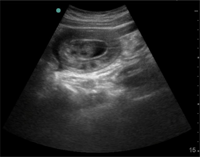
Point of care ultrasound for acute abdomen (ACUTE approach) showed localized free fluid with doughnut sign edematous bowel loop wall

Point of care ultrasound for acute abdomen (ACUTE approach) showed bowel telescoping inside itself with edematous bowel loop wall (long axis)
Abdominal X-ray was normal
After ultrasound finding, we decided to perform urgent CT scan of the abdomen which showed the evidence of long segment entero-enteric (jejuno-jejunal) intussusception seen mainly in the left side of the abdomen, with relatively collapsed intussusceptum segment. Mild amount of intraperitoneal free fluid was seen in the jejunal mesentery next to the involved segments as well as in the pelvic peritoneal pouches (Fig. 3 ).
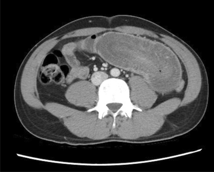
CT scan abdomen with contrast showed a long segment entero-enteric (jejuno-jejunal) intussusception seen mainly in the left side of the abdomen
Surgical consultation was obtained; however, the patient preferred to continue the medical care in his country.
A 70-year-old female diabetic and hypertensive was presented to Rashid Emergency and Trauma Center with generalized abdominal pain for 3 h after food which was associated with vomiting and diarrhea twice. There was no urinary complaint, chest pain or fever.
Vital signs were normal. Abdomen was soft with mild tenderness in the right lower abdomen, without peritonitis clinical sign. Laboratory test was insignificant, except for leukocytosis 17.2·10 3 /μL (3.6–11.0·10 3 /μL) and plasma lactic acid 3.7 mmol/L (0.5–2.2 mmol/L).
C: normal IVC
T: localized free fluid with dilated small bowel loop in right lower quadrant with absent peristalsis (Fig. 4 ).

Point of care ultrasound for acute abdomen showed localized free fluid * with dilated small bowel loop in right lower quadrant with absent peristalsis
CT abdomen showed evidence of significantly dilated small bowel loops in the right side of the lower abdomen and pelvis with a short segment critical stenosis measuring about 2.3 cm. It also showed an evidence suggestive of internal herniation with dilated loops of bowel. Jejunal loops and distal ileum appear collapsed. Free fluid was seen in the abdomen and pelvis (Fig. 5 ).
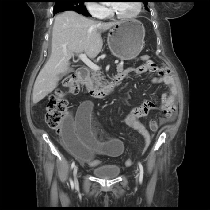
CT abdomen with contrast showed dilated small bowel loops in the right side of the lower abdomen and pelvis with a short segment critical stenosis measuring about 2.3 cm
Urgent surgical consultation was obtained and the patient had undergone laparotomy which was conclusive of an omental band adherent to the small bowel mesentery.
About 150 cm proximal to ileo cecal juction producing congestion with punctuate haemorrhages and dilatation of the proximal bowel of about 30 cm. After the band was released; the color of the bowel changed to normal with good peristalsis. No bowel resection was needed.
Patient post-operative course was uneventful, and she was discharged home after 5 days.
Patient symptoms that mimicked simple gastroenteritis had improved, but because of our ultrasound finding that suggested intestinal obstruction, it was decided to perform a CT abdomen. If not for our preliminary ultrasound findings, this patient would have been discharged without further imaging.
POCUS approaches have been published in multiple critical care conditions such as Bedside Lung Ultrasound in Emergency (BLUE) protocol in respiratory failure which concluded that lung ultrasound immediately provided the diagnosis of acute respiratory failure in 90.5% of cases. The BLUE protocol focused on lung ultrasound only [ 4 ]; however, the RUSH protocol demonstrated that initial integration of bedside ultrasound into the evaluation of the patient with shock results in a more accurate initial diagnosis with an improved patient care plan [ 5 ].
In this case series, we presented two cases where ACUTE ABDOMEN played an important role in their management, POCUS can play an important role in acute abdomen, by early recognition and diagnosing critical conditions. Instead of relying only on physical examination which might have very low sensitivity in comparison to ultrasound, for example in physical examination can identify 38% of patients with AAA [ 6 ], ultrasound by emergency physician can detect AAA in 93% to 100% of cases, with specificities approaching 100% [ 7 , 8 , 9 ].
In other pathologies like pneumoperitoneum, X-ray is more frequently used, but ultrasound is superior to upright chest and lateral decubitus X-ray where the sensitivity is 92% versus 78% for X-ray [ 10 ].
The two cases presented with undifferentiated acute abdomen, in which bedside ultrasound was started with ACUTE approach after history and physical examination, (Table 1 A) [ 11 ] showed unexpected finding, which needed further radiological evaluation by CT scan with contrast and early referral to surgical team.
For the second part of the approach, ABDOMEN (Table 1 B) [ 11 ] usually will be a secondary survey and will focus the assessment with guide of the history and examination.
Conclusions
ACUTE and ABDOMEN sonographic approach in acute abdomen can play an important role in ruling out critical diagnosis, and can guide emergency physician or any critical care physician in patient management.
Further prospective studies are warranted.
Availability of data and materials
Not applicable.
Abbreviations
point of care ultrasound
bedside lung ultrasound in emergency
rapid ultrasound in shock
abdominal aortic aneurysm
inferior vena cava
Pathan S, Mitra B, Mirza S, Momin U, Ahmed Z, Andraous L et al (2018) Emergency physician interpretation of point-of-care ultrasound for identifying and grading of hydronephrosis in renal colic compared with consensus interpretation by emergency radiologists. Acad Emerg Med 25(10):1129–1137
Article Google Scholar
Ultrasound Guidelines: emergency, point-of -care, and clinical ultrasound guidelines in medicine. Acep.org (2019). https://acep.org/patient-care/policy-statements/ultrasound-guidelines-emergency-point-of–care-andclinical-ultrasound-guidelines-in-medicine/ . Accessed 14 Mar 2019
Abdominal Pain in ED (2017) Using a novel sonographic approach Maryam Al Ali and Salma Alrajaby Emergency Department, Rashid Hospital, Dubai, United Arab Emirates. Eur J Emerg Med. 24:e1–e15
Google Scholar
Lichtenstein D, Mezière G (2008) Relevance of lung ultrasound in the diagnosis of acute respiratory failure: the BLUE Protocol. Chest 134(1):117–125
Perera P, Mailhot T, Riley D, Mandavia D (2012) The RUSH Exam 2012: rapid ultrasound in shock in the evaluation of the critically ill patient. Ultrasound Clin 7(2):255–278
Chervu A, Clagett G, Valentine R, Myers S, Rossi P (1995) Role of physical examination in detection of abdominal aortic aneurysms. Surgery 117(4):454–457
Article CAS Google Scholar
Dent B, Kendall RJ, Boyle AA et al (2007) Emergency ultrasound of the abdominal aorta by UK emergency physicians: a prospective cohort study. Emerg Med J 24:547–549
Costantino TG, Bruno EC, Handly N et al (2005) Accuracy of emergency medicine ultrasound in the evaluation of abdominal aortic aneurysm. J Emerg Med 29:455–460
Tayal VS, Graf CD, Gibbs MA (2003) Prospective study of accuracy and outcome of emergency ultrasound for abdominal aortic aneurysm over two years. Acad Emerg Med 10:867–871
Chen S, Yen Z, Wang H, Lin F, Hsu C, Chen W (2002) Ultrasonography is superior to plain radiography in the diagnosis of pneumoperitoneum. Br J Surg 89(3):351–354
Case Report: POCUS to FOCUS–POCUS Journal. Pocusjournal.com (2018). http://pocusjournal.com/article/2018-03-01p15-17/ . Accessed 28 Aug 2018
Download references
Acknowledgements
Author information, authors and affiliations.
Arab Board in Emergency Medicine, Rashid Hospital Trauma Center, Dubai Health Authority, oud metha, Dubai, United Arab Emirates
Maryam Al Ali & Salma Alrajaby
Emergency Medicine Resident, Rashid Hospital Trauma Center, Dubai Health Authority, oud metha, Dubai, United Arab Emirates
Sarah Jabbour
You can also search for this author in PubMed Google Scholar
Contributions
SJ: presentation of the cases, MAA and SA: discussion of the case. All authors read and approved the final manuscript.
Corresponding author
Correspondence to Maryam Al Ali .
Ethics declarations
Ethics approval and consent to participate, consent for publication, competing interests.
The authors declare that they have no competing interests.
Additional information
Publisher's note.
Springer Nature remains neutral with regard to jurisdictional claims in published maps and institutional affiliations.
Rights and permissions
Open Access This article is distributed under the terms of the Creative Commons Attribution 4.0 International License ( http://creativecommons.org/licenses/by/4.0/ ), which permits unrestricted use, distribution, and reproduction in any medium, provided you give appropriate credit to the original author(s) and the source, provide a link to the Creative Commons license, and indicate if changes were made.
Reprints and permissions
About this article
Cite this article.
Al Ali, M., Jabbour, S. & Alrajaby, S. ACUTE ABDOMEN systemic sonographic approach to acute abdomen in emergency department: a case series. Ultrasound J 11 , 22 (2019). https://doi.org/10.1186/s13089-019-0136-5
Download citation
Received : 20 November 2018
Accepted : 24 August 2019
Published : 23 September 2019
DOI : https://doi.org/10.1186/s13089-019-0136-5
Share this article
Anyone you share the following link with will be able to read this content:
Sorry, a shareable link is not currently available for this article.
Provided by the Springer Nature SharedIt content-sharing initiative
- Acute abdomen
- Emergency ultrasound
- Critical ultrasound

Presentations made painless
- Get Premium
106 Ultrasound Essay Topic Ideas & Examples
Inside This Article
Ultrasound technology has revolutionized the field of medicine, allowing healthcare professionals to visualize internal structures and organs without invasive procedures. As a result, ultrasound has become an essential tool for diagnosing and monitoring various medical conditions. If you are a student studying ultrasound technology or a healthcare professional looking to expand your knowledge, here are 106 ultrasound essay topic ideas and examples to help you explore this fascinating field further.
- The history and development of ultrasound technology
- The physics behind ultrasound imaging
- The role of ultrasound in obstetrics and gynecology
- Ultrasound-guided procedures in interventional radiology
- The use of ultrasound in diagnosing musculoskeletal injuries
- Ultrasound imaging of the heart (echocardiography)
- The benefits and limitations of 3D/4D ultrasound imaging
- Contrast-enhanced ultrasound for liver imaging
- Ultrasound elastography for assessing tissue stiffness
- The role of ultrasound in diagnosing breast cancer
- Ultrasound imaging in emergency medicine
- Point-of-care ultrasound in critical care settings
- The use of ultrasound in vascular imaging
- Ultrasound-guided nerve blocks for pain management
- The future of ultrasound technology in healthcare
- Ultrasound imaging of the thyroid gland
- The use of ultrasound in diagnosing gallbladder disease
- Ultrasound-guided biopsy procedures
- Ultrasound imaging of the kidneys
- The role of ultrasound in diagnosing appendicitis
- Ultrasound imaging of the pancreas
- The use of ultrasound in diagnosing gastrointestinal disorders
- Ultrasound-guided injections for joint pain
- Ultrasound imaging of the urinary tract
- The benefits of portable ultrasound technology
- Ultrasound imaging of the prostate gland
- The use of ultrasound in diagnosing testicular conditions
- Ultrasound-guided drainage procedures
- Ultrasound imaging of the spleen
- The role of ultrasound in diagnosing hernias
- Ultrasound-guided nerve ablation for pain management
- Ultrasound imaging of the placenta
- The use of ultrasound in diagnosing fetal anomalies
- Ultrasound-guided thyroid biopsy procedures
- Ultrasound imaging of the adrenal glands
- The benefits of contrast-enhanced ultrasound for liver imaging
- Ultrasound-guided joint injections for arthritis
- Ultrasound imaging of the parathyroid glands
- The role of ultrasound in diagnosing lymph node abnormalities
- Ultrasound-guided breast biopsy procedures
- Ultrasound imaging of the thymus gland
- The use of ultrasound in diagnosing mediastinal masses
- Ultrasound-guided pleural procedures
- Ultrasound imaging of the pericardium
- The benefits of contrast-enhanced ultrasound for vascular imaging
- Ultrasound-guided nerve blocks for chronic pain management
- Ultrasound imaging of the carotid arteries
- The role of ultrasound in diagnosing peripheral vascular disease
- Ultrasound-guided varicose vein procedures
- Ultrasound imaging of the aorta
- The use of ultrasound in diagnosing deep vein thrombosis
- Ultrasound-guided sclerotherapy for spider veins
- Ultrasound imaging of the liver and biliary system
- The benefits of contrast-enhanced ultrasound for renal imaging
- Ultrasound-guided renal biopsy procedures
- The role of ultrasound in diagnosing adrenal tumors
- Ultrasound-guided adrenal vein sampling procedures
- Ultrasound imaging of the pancreas and spleen
- The use of ultrasound in diagnosing pancreatic cancer
- Ultrasound-guided pancreatic biopsy procedures
- Ultrasound imaging of the gallbladder and biliary system
- The benefits of contrast-enhanced ultrasound for pancreatic imaging
- Ultrasound-guided percutaneous cholecystostomy procedures
- Ultrasound imaging of the gastrointestinal tract
- The role of ultrasound in diagnosing inflammatory bowel disease
- Ultrasound-guided intestinal biopsy procedures
- Ultrasound imaging of the kidneys and urinary tract
- The use of ultrasound in diagnosing kidney stones
- Ultrasound-guided percutaneous nephrolithotomy procedures
- Ultrasound imaging of the female reproductive system
- The benefits of contrast-enhanced ultrasound for gynecologic imaging
- Ultrasound-guided ovarian cyst aspiration procedures
- Ultrasound imaging of the male reproductive system
- The role of ultrasound in diagnosing testicular cancer
- Ultrasound-guided testicular biopsy procedures
- Ultrasound imaging of the musculoskeletal system
- The use of ultrasound in diagnosing sports injuries
- Ultrasound-guided joint aspiration procedures
- Ultrasound imaging of the nervous system
- The benefits of contrast-enhanced ultrasound for neuroimaging
- Ultrasound-guided nerve conduction studies
- Ultrasound imaging of the head and neck
- The role of ultrasound in diagnosing thyroid nodules
- Ultrasound-guided thyroid fine-needle aspiration biopsy procedures
- Ultrasound imaging of the chest and lungs
- The use of ultrasound in diagnosing pleural effusions
- Ultrasound-guided thoracentesis procedures
- Ultrasound imaging of the heart and blood vessels
- The benefits of contrast-enhanced ultrasound for cardiac imaging
- Ultrasound-guided cardiac catheterization procedures
- Ultrasound imaging of the liver and spleen
- The role of ultrasound in diagnosing liver cirrhosis
- Ultrasound-guided liver biopsy procedures
- Ultrasound imaging of the pancreas and biliary system
- The use of ultrasound in diagnosing pancreatic pseudocysts
- Ultrasound-guided percutaneous drainage procedures
- Ultrasound imaging of the gastrointestinal tract and kidneys
- The benefits of contrast-enhanced ultrasound for urologic and gastrointestinal imaging
- Ultrasound-guided percutaneous nephrostomy procedures
- Ultrasound imaging of the female reproductive system and bladder
- The role of ultrasound in diagnosing pelvic organ prolapse
- Ultrasound-guided bladder sling procedures
- Ultrasound imaging of the male reproductive system and prostate
- The use of ultrasound in diagnosing benign prostatic hyperplasia
- Ultrasound-guided prostate biopsy procedures
These essay topic ideas and examples cover a wide range of ultrasound applications and specialties, providing you with ample opportunities to explore and research this exciting field further. Whether you are a student or a healthcare professional, delving into these topics can deepen your understanding of ultrasound technology and its role in modern medicine.
Want to research companies faster?
Instantly access industry insights
Let PitchGrade do this for me
Leverage powerful AI research capabilities
We will create your text and designs for you. Sit back and relax while we do the work.
Explore More Content
- Privacy Policy
- Terms of Service
© 2024 Pitchgrade
An official website of the United States government
The .gov means it’s official. Federal government websites often end in .gov or .mil. Before sharing sensitive information, make sure you’re on a federal government site.
The site is secure. The https:// ensures that you are connecting to the official website and that any information you provide is encrypted and transmitted securely.
- Publications
- Account settings
Preview improvements coming to the PMC website in October 2024. Learn More or Try it out now .
- Advanced Search
- Journal List
- Ultrasonography
- v.39(2); 2020 Apr
Role of ultrasound in the evaluation of first-trimester pregnancies in the acute setting
Venkatesh a. murugan.
Department of Radiology, University of Massachusetts Medical School, Worcester, MA, USA
Bryan O’Sullivan Murphy
Carolyn dupuis, alan goldstein, young h. kim.
In patients presenting for an evaluation of pregnancy in the first trimester, transvaginal ultrasound is the modality of choice for establishing the presence of an intrauterine pregnancy; evaluating pregnancy viability, gestational age, and multiplicity; detecting pregnancy-related complications; and diagnosing ectopic pregnancy. In this pictorial review article, the sonographic appearance of a normal intrauterine gestation and the most common complications of pregnancy in the first trimester in the acute setting are discussed.

Introduction
The first trimester of pregnancy consists of the first 12-13 weeks, calculated as beginning on the first date of the last menstrual period (LMP). During the first trimester, transvaginal ultrasonography (TVUS) is the imaging modality of choice for both diagnosis and imaging follow-up. The advantages of ultrasound imaging include its widespread availability, relatively low cost, and the acquisition of real-time, high-resolution images. The initial diagnosis of pregnancy is usually made by identifying the presence of serum beta-human chorionic gonadotropin (β-hCG). Ultrasound is then utilized during the first and second trimesters to establish the gestational age of the pregnancy and eventually to evaluate fetal anatomy. In the first trimester, pelvic ultrasound is employed to establish the presence or absence of an intrauterine gestational sac and to evaluate the viability of the pregnancy. In addition, it can be used to evaluate ectopic pregnancy and other pregnancy-related complications. Practice parameters for the performance and recording of obstetric ultrasound images have been described by the American Institute of Ultrasound in Medicine [ 1 ].
Timeline of First-Trimester Sonographic Findings
Gestational sac.
Conventionally, gestational age is initially calculated from the first day of the LMP. Ovulation typically occurs mid-cycle, at about day 14 of the menstrual cycle, at which point fertilization (conception) is most likely to occur. Thus, by the time of the first missed menstrual period, fertilization and implantation of the fertilized ovum have occurred. During the first 3 weeks following conception, the developing gestational sac is below the limit of detection by TVUS [ 2 ]. The growth rate of the gestational sac is approximately 1.1 mm/day and the gestational sac first becomes apparent on TVUS at approximately 4.5-5 weeks of gestational age, appearing as a round anechoic structure located eccentrically within the echogenic decidua ( Table 1 , Fig. 1A , ,B) B ) [ 3 ].

A. Transvaginal ultrasonography (TVUS) demonstrates an anechoic structure with peripheral echogenic tissue (arrowheads) representing a gestational sac in the uterine cavity of a woman with a positive urine pregnancy test. B. TVUS shows a circumscribed heterogenous structure with peripheral vascularity in the right ovary compatible with a corpus luteum (arrowheads).
Timeline of normal fetal development
| Time (wk) | Development milestone |
|---|---|
| 4.5-5 | Appearance of gestational sac |
| 5-5.5 | Yolk sac becomes apparent |
| 6 | Embryo is seen, cardiac pulsation |
| 6.7-7 | Amniotic membrane appears |
| 7-8 | Appearance of fetal spine |
| 8 | Head and limbs begin to appear as distinct from torso |
| 8-8.5 | Fetal motion is appreciable |
| 8-10 | Rhombencephalon appears |
Double Decidual Sac Sign
Subsequent to the appearance of the gestation sac, two concentric echogenic rings encircling the central anechoic collection develop: the outer ring represents the decidua parietalis, while the inner ring represents the decidua capsularis and chorion ( Fig. 2A , ,B). B ). This is known as the double decidual sac sign (DDS), which is a definitive sign of an intrauterine pregnancy (IUP). While the presence of the DDS sign confirms an IUP, its absence does not exclude an IUP [ 4 ]. Furthermore, the DDS sign can be difficult to demonstrate sonographically. For this reason, the possibility of a gestational sac should be considered for any round or ovoid fluid collection within the endometrium ( Fig. 3A , ,B) B ) [ 4 ]. Gestational sac size is measured in 3 dimensions and the mean sac diameter (MSD) is used to help estimate the early gestational age.

A. Transvaginal ultrasonography demonstrates two concentric echogenic rings (arrowheads) with intervening trace hypoechoic material, known as the double decidual sac sign. B. Graphical representation of the double decidual sign is shown.

A. Initial transvaginal ultrasonography (TVUS) shows a vague hypoechoic collection measuring 7 mm in the uterine fundus (arrow). The morphology was not typical for an intrauterine pregnancy. B. Subsequent TVUS 2 weeks later demonstrates an intrauterine gestational sac with an embryo with heart rate of 126 bpm.
At around 5.5 weeks of gestation, the developing yolk sac becomes visible. Initially appearing as two echogenic parallel lines at the periphery of the gestational sac, the yolk sac eventually acquires its typical round appearance by the end of 5.5 weeks [ 2 ].
The embryo (sometimes referred to as the fetal pole early on) becomes apparent at 6 weeks of gestation as a relatively featureless echogenic linear or oval structure adjacent to the yolk sac, initially measuring 1-2 mm in length. At this point, the MSD is approximately 10 mm. The crown-rump length (CRL) is the measurement between the cranial and caudal ends of the embryo and is the most accurate measure of the gestational age in the first trimester. The CRL gradually increases, measuring 10 mm at 7.0 weeks. The lack of a visible embryo on TVUS once the MSD reaches at least 25 mm is diagnostic of pregnancy failure ( Fig. 4 ) [ 2 , 5 ]. While the fetal pole begins as a featureless structure, some fetal anatomic structures become visible as the first-trimester progresses. The spine appears at 7-8 weeks, and the hindbrain (rhombencephalon) is evident at 8-10 weeks [ 6 ].

A. Initial ultrasonography shows a gestational sac without a yolk sac or embryo. B. Follow-up ultrasonography 2 weeks later shows a gestational sac measuring greater than 25 mm in diameter without evidence of a yolk sac or embryo. These findings are diagnostic of early pregnancy loss.
Amniotic Membrane
The amniotic membrane becomes visible around 7 weeks, and the CRL closely corresponds to the amniotic sac diameter between 6.5 and 10 weeks of gestation ( Fig. 5 ) [ 7 ]. After fetal urine production commences at about 10 weeks, there is a disproportionate enlargement of the amniotic sac relative to the chorionic cavity. The amnion and chorion fuse after the first trimester at 14-16 weeks [ 8 ].

A curvilinear echogenic membrane is noted around the embryo, corresponding to the amniotic membrane (arrow).
Cardiac Activity
Cardiac activity is seen as early as the sixth week of gestation, when the embryo is 1-2 mm in size. The current guidelines of the Society of Radiologists in Ultrasound (SRU) establish a CRL cutoff of 7 mm, above which one should definitively visualize fetal cardiac activity. The absence of a detectable heartbeat once the embryo measures greater than 7 mm in length is diagnostic of pregnancy failure ( Fig. 6A , ,B) B ) [ 7 ]. The fetal heart rate gradually increases with gestational age from approximately 110 beats per minute (bpm) at 6.2 weeks to approximately 159 bpm at 7.6-8.0 weeks ( Fig. 7A-E ) [ 9 ]. Slow embryonic heart rates are associated with a worse shortterm prognosis, with fetal heart rates less than 100 bpm before 6.3 weeks or below 120 bpm at 6.3-7.0 weeks linked to an increased rate of embryonic demise [ 9 , 10 ]. The overall prognosis improves with increasing heart rate [ 10 ].

A. Transvaginal ultrasonography shows an intrauterine pregnancy with an embryo (arrow) with a crown-rump length of 1.1 cm, corresponding to a gestational age of 7 weeks, 2 days. B. No fetal heart rate was identified, compatible with intrauterine embryonic demise.

A-E. M-mode images show progressive increase in heart rate with advancing gestational age. A. Intrauterine gestation with an embryo and yolk sac (crown-rump length of 7 mm --> gestational age [GA] of 6 weeks and 5 days) is shown. B. Fetal heart rate is 127 bpm. C. The cervix is closed. D. Two days later, pelvic ultrasonography demonstrates interval growth of the embryo, with a crown-rump length of 1 cm, corresponding to a GA of 7 weeks. E. The heart rate increases progressively with advancing gestational age.
First-Trimester Abnormalities
First-trimester TVUS is routinely performed in patients presenting with pelvic/abdominal pain or vaginal bleeding. Once pregnancy is established with urine or serum β-hCG tests, the utility of TVUS for evaluation of these patients is multifactorial: (1) to determine the presence and multiplicity of an IUP, (2) to determine the viability of an IUP, (3) to determine the stage of spontaneous abortion in the case of a nonviable pregnancy, and (4) to identify probable reasons why an IUP is not identified on TVUS.
Confirming an IUP
The detection of an eccentrically-located, anechoic collection in the endometrium of a patient with elevated serum β-hCG levels represents an IUP in 99.5% of cases [ 4 ]. The presence of two or more gestational sacs surrounded by thick echogenic chorion, or sonographic features of the inter-twin membrane and "twin-peak" sign, confirm a multiple-gestation pregnancy [ 11 ].
Evaluating Viability
Once an IUP is identified, the viability and presence or absence of abnormal features must be evaluated. The timeline of visualization of the gestational sac, yolk sac, and embryo at 5, 5.5, and 6 weeks, respectively, are accurate and consistent [ 5 ]. Deviations from the normal chronological appearance of these structures are highly suspicious for pregnancy failure. The SRU has presented specific guidelines for diagnosing pregnancy failure based on certain characteristics: namely, (1) the CRL measurement by which an embryonic heart rate must be identified (7 mm), (2) the MSD by which an embryo should be identified (25 mm), and (3) the absence of an embryo in two consecutive ultrasound exams separated by a fixed time interval. In addition, other findings including the empty amnion sign, a yolk sac greater than 7 mm, and a disproportionately small gestational sac are highly suspicious for pregnancy failure ( Fig. 8 ). Through these guidelines, the SRU aims to achieve 100% specificity for defining pregnancy failure and to sustain a primum non nocere approach given the calamitous outcome of a potentially normal pregnancy following treatment for an incorrectly diagnosed pregnancy failure ( Table 2 ) [ 4 , 5 ].

Transabdominal ultrasonography in a 34-year-old woman with a positive beta-human chorionic gonadotropin test and vaginal bleeding demonstrates an intrauterine gestation with a mean sac diameter of 23 mm and a yolk sac diameter of 19 mm. No definite fetal pole was identified. Instead, an amorphous embryonic structure (arrowheads) was identified. These findings are suspicious for, but not diagnostic of pregnancy failure.
Findings diagnostic of and suspicious for pregnancy failure
| Finding | Diagnostic of pregnancy failure | Suspicious for pregnancy failure |
|---|---|---|
| Absent fetal cardiac activity by the time CRL is a certain size | CRL ≥7 mm | CRL <7 mm |
| Absent embryo by the time the gestational sac is a certain size | MSD ≥25 mm | MSD 16-24 mm |
| Absent embryo in two consecutive exams separated by time | Nonvisualization of an embryo with fetal heart rate 2 wk after identification of gestational sac without yolk sac | Nonvisualization of an embryo with fetal heart rate 7-10 days after US showed gestational sac with yolk sac |
| Nonvisualization of an embryo with fetal heart rate 11 or more days after identification of a gestational sac with yolk sac | Nonvisualization of embryo 6 wk after LMP | |
| Abnormal morphology of the gestational sac, amnion, and yolk sac | - | Amnion seen adjacent to yolk sac with no visible embryo (empty amnion) |
| - | Yolk sac >7 mm | |
| - | Disproportionately small gestational sac (in relation to size of embryo, <5 mm difference in size between MSD and CRL) |
CRL, crown-rump length; MSD, mean sac diameter; US, ultrasonography; LMP, last menstrual period.
Subchorionic Hematoma
Subchorionic hematoma (SCH) is a relatively common finding in the first trimester and has been reported to occur in 18%-22% of IUPs in patients presenting with vaginal bleeding [ 12 , 13 ]. On TVUS, SCH appears as a crescent-shaped, heterogeneous avascular collection between the gestational sac and decidua basalis ( Fig. 9A , ,B). B ). Larger subchorionic hematomas are associated with an increased risk of pregnancy loss, especially if the hematoma is greater than twothirds of the chorionic circumference [ 13 , 14 ].

A. Transvaginal ultrasonography in a pregnant woman shows a gestational sac with an embryo and a heterogeneous subchorionic collection (arrowheads) encircling approximately 180° of the gestational sac. B. Graphic depiction of the findings in A is shown.
Spontaneous Abortion
Spontaneous abortion or miscarriage is clinically defined as the loss of a pregnancy before the 20th week of gestation or the expulsion of a fetus weighing less than 500 g [ 15 , 16 ]. There are various stages of spontaneous abortion. A threatened abortion refers to a clinical scenario in which a patient presents with vaginal spotting/bleeding and cramping/contractions with a closed cervical os. The pregnancy itself may appear normal or may demonstrate abnormal features. Poor prognostic indicators include abnormal morphology (e.g., a small or irregular gestational sac), fetal bradycardia, or a large SCH [ 9 , 12 ]. An inevitable abortion involves a similar clinical situation with vaginal bleeding and abdominal cramping, but with an open cervical os on TVUS ( Fig. 10A-C ). The products of conception may be normally or abnormally positioned within the uterus or may protrude into the cervix. An incomplete abortion is the term used when the retained products of conception remain within the uterus after passage of the pregnancy. This often appears as a heterogeneous collection or mass within the uterus. While it may be avascular, the presence of blood flow enables the diagnosis of retained products. A completed abortion is the cessation of vaginal bleeding following the passage of the pregnancy without retained products of conception ( Fig. 11A-D ). Lastly, a missed abortion is a nonviable pregnancy with a closed cervix and no clinical symptoms of miscarriage [ 17 ].

A. Initial TVUS shows an intrauterine gestation (arrow), with an open cervix (arrowheads). B. No heart rate was identified. C. Followup ultrasonography obtained the next day shows the gestational sac in the cervical canal (arrowheads), compatible with inevitable abortion.

A, B. Gestational sac containing a fetal pole was identified in the cervix (arrow); the abortion was in progress. C. No fetal heart rate was identified. D. The patient passed a few clots and transvaginal images were obtained. The previously seen gestational sac in the cervix was no longer seen, compatible with a completed abortion.
Pregnancy of Unknown Location and Ectopic Pregnancy
In a substantial number of patients evaluated in the emergency department during very early pregnancy, the location of the gestational sac is inconclusive. The significance of a nonvisualized gestational sac on TVUS in a patient with a positive pregnancy test could reflect one of three scenarios: (1) less than 5 weeks of gestation, (2) ectopic pregnancy, or (3) a completed abortion [ 18 ].
It is incumbent for the technologist/radiologist to carefully scrutinize the adnexa and other spaces in the pelvis for any masses, collections, or obvious products of conception in order to rule out an ectopic pregnancy. As the prevalence of ectopic pregnancy is 1.4% and it accounts for 25% of all maternal deaths, practitioners should have a high degree of suspicion for this diagnosis. Although the vast majority of ectopic pregnancies occur in the fallopian tubes, implantation of pregnancies at other sites can also take place, including the cervix, cesarean scars, uterine cornua, and other nongynecological sites in the abdomen and pelvis ( Figs. 12 - - 14) 14 ) [ 2 ]. In cases in which no IUP is identified and there is no sonographic evidence of an ectopic pregnancy, serial monitoring of β-hCG levels and short-term repeat TVUS are generally recommended for follow-up.

A. No intrauterine gestational sac was identified. The right ovary and adnexa were normal. B. A left adnexal heterogenous vascular mass (arrowheads), was suspicious for an adnexal ectopic pregnancy, which was confirmed intraoperatively.

A. No intrauterine gestational sac was identified. The left ovary and adnexa were normal. B, C. Sonograms demonstrate a right adnexal mass containing a gestational sac (arrowheads) and a fetal pole (arrow), with a heart rate of 167 bpm, compatible with a right adnexal ectopic gestation.

A. Transabdominal ultrasonography in a woman with a positive beta-human chorionic gonadotropin test, shows a gestational sac containing a fetal pole in the cervix (arrow). B. Transvaginal ultrasonography shows a gestational sac containing a yolk sac (arrowheads) and fetal pole (arrow).
Gestational Trophoblastic Disease
Gestational trophoblastic disease is a broad term which encompasses both benign entities, such as partial and complete mole, gestational trophoblastic neoplasia (GTN), and malignant diagnoses, such as invasive mole, choriocarcinoma, and epithelioid and placental site trophoblastic tumors. As a result of dispermic fertilization of an ovum, pregnant patients will often present with vaginal bleeding. On TVUS in the first trimester, the endometrial cavity will contain an echogenic solid mass, usually with numerous cystic spaces, which are the hydropic villi and trophoblastic hyperplasia ( Fig. 15 ). Careful scrutiny of the mass is important to distinguish between complete mole (no fetal parts), partial mole (some fetal parts), and GTN (myometrial invasion) [ 19 ].

Transvaginal ultrasonography in a 35-year-old woman presenting with an elevated serum beta-human chorionic gonadotropin (β -hCG) level (>383,000 mIU/mL) and vaginal bleeding, shows an echogenic, heterogenous mass with minimal peripheral vascularity (arrowheads) and numerous cystic spaces. No fetal parts or myometrial invasion was identified. These findings, given the significantly elevated β-hCG value, were diagnostic of complete mole.
The diagnostic possibilities for pregnant patients presenting with pain and bleeding are broad. TVUS is paramount in its utility as a diagnostic tool for these patients. When used in combination with clinical information and serum β-hCG levels, it can provide diagnostic and prognostic information to clinicians regarding pregnancy confirmation and viability, as well as rapid information regarding life-threatening conditions such as ectopic pregnancy.
Author Contributions
Conceptualization: Kim YH. Data acquisition: Murugan VA, Murphy BO. Data analysis or interpretation: Murugan VA, Murphy BO. Drafting of manuscript: Murugan VA, Murphy BO. Critical revision of manuscript: Dupuis C, Goldstein A, Kim YH. Approval of the final version of the manuscript: all authors.
No potential conflict of interest relevant to this article was reported.
Open Access is an initiative that aims to make scientific research freely available to all. To date our community has made over 100 million downloads. It’s based on principles of collaboration, unobstructed discovery, and, most importantly, scientific progression. As PhD students, we found it difficult to access the research we needed, so we decided to create a new Open Access publisher that levels the playing field for scientists across the world. How? By making research easy to access, and puts the academic needs of the researchers before the business interests of publishers.
We are a community of more than 103,000 authors and editors from 3,291 institutions spanning 160 countries, including Nobel Prize winners and some of the world’s most-cited researchers. Publishing on IntechOpen allows authors to earn citations and find new collaborators, meaning more people see your work not only from your own field of study, but from other related fields too.
Brief introduction to this section that descibes Open Access especially from an IntechOpen perspective
Want to get in touch? Contact our London head office or media team here
Our team is growing all the time, so we’re always on the lookout for smart people who want to help us reshape the world of scientific publishing.
Home > Books > Medical Imaging
Ultrasound Imaging - Current Topics

Book metrics overview
4,529 Chapter Downloads
Impact of this book and its chapters
Total Chapter Downloads on intechopen.com
Book Citations
Total Chapter Citations
Academic Editor
Gregory University , Nigeria
Published 11 May 2022
Doi 10.5772/intechopen.95178
ISBN 978-1-78985-186-1
Print ISBN 978-1-78984-877-9
eBook (PDF) ISBN 978-1-78985-331-5
Copyright year 2022
Number of pages 154
Ultrasound Imaging - Current Topics presents complex and current topics in ultrasound imaging in a simplified format. It is easy to read and exemplifies the range of experiences of each contributing author. Chapters address such topics as anatomy and dimensional variations, pediatric gastrointestinal emergencies, musculoskeletal and nerve imaging as well as molecular sonography. The book is...
Ultrasound Imaging - Current Topics presents complex and current topics in ultrasound imaging in a simplified format. It is easy to read and exemplifies the range of experiences of each contributing author. Chapters address such topics as anatomy and dimensional variations, pediatric gastrointestinal emergencies, musculoskeletal and nerve imaging as well as molecular sonography. The book is a useful resource for researchers, students, clinicians, and sonographers looking for additional information on ultrasound imaging beyond the basics.
By submitting the form you agree to IntechOpen using your personal information in order to fulfil your library recommendation. In line with our privacy policy we won’t share your details with any third parties and will discard any personal information provided immediately after the recommended institution details are received. For further information on how we protect and process your personal information, please refer to our privacy policy .
Cite this book
There are two ways to cite this book:
Edited Volume and chapters are indexed in
Table of contents.
By Solomon Demissie, Mulatie Atalay and Yonas Derso
By Ercan Ayaz
By Haithem Zaafouri, Meryam Mesbahi, Nizar Khedhiri, Wassim Riahi, Mouna Cherif, Dhafer Haddad and Anis Ben Maamer
By Felix Okechukwu Erondu
By Stefan Cristian Dinescu, Razvan Adrian Ionescu, Horatiu Valeriu Popoviciu, Claudiu Avram and Florentin Ananu Vreju
By María Eugenia Aponte-Rueda and María Isabel de Abreu
By Jong Hwa Lee, Jae Uk Lee and Seung Wan Yoo
By J.M. López Álvarez, O. Pérez Quevedo, S. Alonso-Graña López-Manteola, J. Naya Esteban, J.F. Loro Ferrer and D.L. Lorenzo Villegas
By Arthur Fleischer and Sai Chennupati
IMPACT OF THIS BOOK AND ITS CHAPTERS
4,529 Total Chapter Downloads
2 Crossref Citations
3 Dimensions Citations
Order a print copy of this book
Hardcover | Printed Full Colour
Available on

Delivered by

£119 (ex. VAT)*
FREE SHIPPING WORLDWIDE
* Residents of European Union countries need to add a Book Value-Added Tax Rate based on their country of residence. Institutions and companies, registered as VAT taxable entities in their own EU member state, will not pay VAT by providing IntechOpen with their VAT registration number. This is made possible by the EU reverse charge method.
Related books
Medical imaging.
Edited by Felix Okechukwu Erondu
Medical Imaging in Clinical Practice
Medical and biological image analysis.
Edited by Robert Koprowski
Edited by Yongxia Zhou
Ultrasound Elastography
Edited by Monica Lupsor Platon
New Advances in Magnetic Resonance Imaging
Edited by Denis Larrivee
Frontiers in Neuroimaging
Edited by Xianli Lv
Optical Coherence Tomography
Edited by Giuseppe Lo Giudice
Updates in Endoscopy
Edited by Somchai Amornyotin
Elastography
Edited by Dana Stoian
Call for authors
Submit your work to intechopen.

IntechOpen Author/Editor? To get your discount, log in .
Discounts available on purchase of multiple copies. View rates
Local taxes (VAT) are calculated in later steps, if applicable.
Support: [email protected]
- Patient Care & Health Information
- Tests & Procedures
Diagnostic ultrasounds use sound waves to make pictures of the body. Ultrasound, also called sonography, shows the structures inside the body. The images can help guide diagnosis and treatment for many diseases and conditions.
Most ultrasounds are done using a device outside the body. However, some involve placing a small device inside the body.
Products & Services
- A Book: Mayo Clinic Family Health Book
- Assortment of Products for Daily Living from Mayo Clinic Store
- Newsletter: Mayo Clinic Health Letter — Digital Edition
Why it's done
Ultrasound is used for many reasons, including to:
- View the uterus and ovaries during pregnancy and monitor the developing baby's health.
- Diagnose gallbladder disease.
- Evaluate blood flow.
- Guide a needle for biopsy or tumor treatment.
- Examine a breast lump.
- Check the thyroid gland.
- Find genital and prostate problems.
- Assess joint inflammation, called synovitis.
- Evaluate metabolic bone disease.
More Information
- Abdominal aortic aneurysm
- Acute kidney injury
- Acute liver failure
- Acute lymphocytic leukemia
- Adenomyosis
- Adult Still disease
- Alcoholic hepatitis
- Anal cancer
- Appendicitis
- Arteriosclerosis / atherosclerosis
- Arteriovenous fistula
- Atelectasis
- Atypical genitalia
- Autonomic neuropathy
- Bladder stones
- Blood in urine (hematuria)
- Breast cancer
- Breast pain
- Carotid artery disease
- Cerebral palsy
- Cholestasis of pregnancy
- Chronic exertional compartment syndrome
- Chronic kidney disease
- Cleft lip and cleft palate
- Congenital adrenal hyperplasia
- Conjoined twins
- Deep vein thrombosis (DVT)
- Double uterus
- Down syndrome
- Ductal carcinoma in situ (DCIS)
- Endometrial cancer
- Endometriosis
- Enlarged breasts in men (gynecomastia)
- Enlarged liver
- Epididymitis
- Erectile dysfunction
- Eye melanoma
- Fibroadenoma
- Fibrocystic breasts
- Galactorrhea
- Ganglion cyst
- Glomerulonephritis
- Growth plate fractures
- Hamstring injury
- High blood pressure in children
- Hurthle cell cancer
- Incompetent cervix
- Infant reflux
- Inflammatory breast cancer
- Intussusception
- Invasive lobular carcinoma
- Iron deficiency anemia
- Ischemic colitis
- Kidney cancer
- Knee bursitis
- Liver cancer
- Liver disease
- Liver hemangioma
- Male breast cancer
- Mammary duct ectasia
- Median arcuate ligament syndrome (MALS)
- Menstrual cramps
- Miscarriage
- Morning sickness
- Morton's neuroma
- Multisystem inflammatory syndrome in children (MIS-C)
- Muscle strains
- Muscular dystrophy
- Myelofibrosis
- Neuroblastoma
- Nonalcoholic fatty liver disease
- Osteoporosis
- Ovarian cancer
- Ovarian cysts
- Painful intercourse (dyspareunia)
- Pancreatic cancer
- Patellar tendinitis
- Pelvic inflammatory disease (PID)
- Peripheral artery disease (PAD)
- Peyronie disease
- Placenta previa
- Placental abruption
- Polycystic kidney disease
- Polymyalgia rheumatica
- Post-vasectomy pain syndrome
- Precocious puberty
- Premature birth
- Preterm labor
- Prostate cancer
- Pulmonary embolism
- Pyloric stenosis
- Recurrent breast cancer
- Residual limb pain
- Retinal detachment
- Retinoblastoma
- Rotator cuff injury
- Sacral dimple
- Sacroiliitis
- Scrotal masses
- Secondary hypertension
- Solitary rectal ulcer syndrome
- Spermatocele
- Spina bifida
- Swollen knee
- Takayasu's arteritis
- Tapeworm infection
- Testicular cancer
- Thrombophlebitis
- Thyroid cancer
- Thyroid nodules
- Torn meniscus
- Toxic hepatitis
- Toxoplasmosis
- Tricuspid atresia
- Tuberous sclerosis
- Uterine fibroids
- Uterine prolapse
- Wilms tumor
- Zollinger-Ellison syndrome
Diagnostic ultrasound is a safe procedure that uses low-power sound waves. There are no known risks.
Ultrasound is a valuable tool, but it has limitations. Sound waves don't travel well through air or bone. This means ultrasound isn't effective at imaging body parts that have gas in them or are hidden by bone, such as the lungs or head. Ultrasound also may not be able to see objects that are located very deep in the human body. To view these areas, your healthcare professional may order other imaging tests, such as CT or MRI scans or X-rays.
How you prepare
Most ultrasound exams require no preparation. However, there are a few exceptions:
- For some scans, such as a gallbladder ultrasound, your healthcare professional may ask that you not eat or drink for a certain period of time before the exam.
- Other scans, such as a pelvic ultrasound, may require a full bladder. Your healthcare professional will let you know how much water you need to drink before the exam. Do not urinate until the exam is done.
- Young children may need additional preparation. When scheduling an ultrasound for yourself or your child, ask your healthcare professional if there are any specific instructions you'll need to follow.
Clothing and personal items
Wear loose clothing to your ultrasound appointment. You may be asked to remove jewelry during your ultrasound. It's a good idea to leave any valuables at home.
What you can expect
Before the procedure.
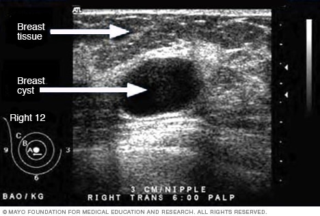
- Ultrasound of breast cyst
This ultrasound shows a breast cyst.
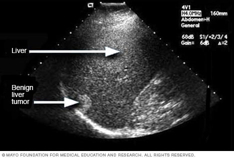
- Ultrasound of liver tumor
An ultrasound uses sound waves to create an image. This ultrasound shows a noncancerous liver tumor.
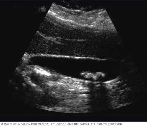
- Ultrasound of gallstones
This ultrasound shows gallstones in the gallbladder.
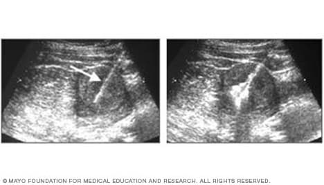
- Ultrasound of needle-guided procedure
These images show how ultrasound can help guide a needle into a tumor (left), where material is injected (right) to destroy tumor cells.
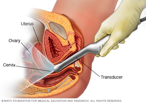
- Transvaginal ultrasound
During a transvaginal ultrasound, a healthcare professional or technician uses a wandlike device called a transducer. The transducer is inserted into your vagina while you lie on your back on an exam table. The transducer emits sound waves that generate images of your pelvic organs.
Before your ultrasound begins, you may be asked to do the following:
- Remove any jewelry from the area being examined.
- Remove or reposition some or all of your clothing.
- Change into a gown.
You'll be asked to lie on an exam table.
During the procedure
Gel is applied to your skin over the area being examined. It helps prevent air pockets, which can block the sound waves that create the images. This safe, water-based gel is easy to remove from skin and, if needed, clothing.
A trained technician, called a sonographer, uses a small, hand-held device called a transducer. The technician presses the transducer against the area being studied and moves it as needed to capture the images. The transducer sends sound waves into your body and collects the ones that bounce back. The images appear on a computer.
Sometimes, ultrasounds are done inside the body. In this case, the transducer is attached to a probe that's inserted into a natural opening in the body. Examples include:
- Transesophageal echocardiogram. A transducer, inserted into the esophagus, obtains heart images. It's usually done while under sedation.
- Transrectal ultrasound. This test creates images of the prostate by placing a special transducer into the rectum.
- Transvaginal ultrasound. A special transducer is inserted into the vagina to look at the uterus and ovaries.
Ultrasound is usually painless. However, you may experience mild discomfort as the sonographer guides the transducer over your body. It may not be comfortable if you're required to have a full bladder or the transducer is inserted it into your body.
A typical ultrasound exam takes from 30 minutes to an hour.
When your exam is complete, a doctor trained to interpret imaging studies, called a radiologist, analyzes the images. The radiologist sends a report to your healthcare professional who will share the results with you.
You should be able to return to usual activities right after an ultrasound.
Clinical trials
Explore Mayo Clinic studies of tests and procedures to help prevent, detect, treat or manage conditions.
- Andreas A, et al., eds. Grainger & Allison's Diagnostic Radiology: A Textbook of Medical Imaging. 7th ed. Elsevier; 2021. https://www.clinicalkey.com. Accessed Jan. 15, 2024.
- General ultrasound. RadiologyInfo.org. https://www.radiologyinfo.org/en/info/genus. Accessed Jan. 15, 2024.
- McKenzie GA (expert opinion). Mayo Clinic. Feb. 1, 2022.
- Chronic pelvic pain
- Ectopic pregnancy
- Fetal macrosomia
- Heavy menstrual bleeding
- Kidney stones
- Molar pregnancy
- Peritonitis
- Rheumatoid arthritis
- Soft tissue sarcoma
- Undescended testicle
- Urinary incontinence
News from Mayo Clinic
- Advancing ultrasound microvessel imaging and AI to improve cancer detection Oct. 13, 2023, 02:01 p.m. CDT
- Doctors & Departments
Mayo Clinic does not endorse companies or products. Advertising revenue supports our not-for-profit mission.
- Opportunities
Mayo Clinic Press
Check out these best-sellers and special offers on books and newsletters from Mayo Clinic Press .
- Mayo Clinic on Incontinence - Mayo Clinic Press Mayo Clinic on Incontinence
- The Essential Diabetes Book - Mayo Clinic Press The Essential Diabetes Book
- Mayo Clinic on Hearing and Balance - Mayo Clinic Press Mayo Clinic on Hearing and Balance
- FREE Mayo Clinic Diet Assessment - Mayo Clinic Press FREE Mayo Clinic Diet Assessment
- Mayo Clinic Health Letter - FREE book - Mayo Clinic Press Mayo Clinic Health Letter - FREE book
5X Challenge
Thanks to generous benefactors, your gift today can have 5X the impact to advance AI innovation at Mayo Clinic.
Have a language expert improve your writing
Run a free plagiarism check in 10 minutes, generate accurate citations for free.
- Knowledge Base
- How to Write a Thesis Statement | 4 Steps & Examples
How to Write a Thesis Statement | 4 Steps & Examples
Published on January 11, 2019 by Shona McCombes . Revised on August 15, 2023 by Eoghan Ryan.
A thesis statement is a sentence that sums up the central point of your paper or essay . It usually comes near the end of your introduction .
Your thesis will look a bit different depending on the type of essay you’re writing. But the thesis statement should always clearly state the main idea you want to get across. Everything else in your essay should relate back to this idea.
You can write your thesis statement by following four simple steps:
- Start with a question
- Write your initial answer
- Develop your answer
- Refine your thesis statement
Instantly correct all language mistakes in your text
Upload your document to correct all your mistakes in minutes

Table of contents
What is a thesis statement, placement of the thesis statement, step 1: start with a question, step 2: write your initial answer, step 3: develop your answer, step 4: refine your thesis statement, types of thesis statements, other interesting articles, frequently asked questions about thesis statements.
A thesis statement summarizes the central points of your essay. It is a signpost telling the reader what the essay will argue and why.
The best thesis statements are:
- Concise: A good thesis statement is short and sweet—don’t use more words than necessary. State your point clearly and directly in one or two sentences.
- Contentious: Your thesis shouldn’t be a simple statement of fact that everyone already knows. A good thesis statement is a claim that requires further evidence or analysis to back it up.
- Coherent: Everything mentioned in your thesis statement must be supported and explained in the rest of your paper.
Here's why students love Scribbr's proofreading services
Discover proofreading & editing
The thesis statement generally appears at the end of your essay introduction or research paper introduction .
The spread of the internet has had a world-changing effect, not least on the world of education. The use of the internet in academic contexts and among young people more generally is hotly debated. For many who did not grow up with this technology, its effects seem alarming and potentially harmful. This concern, while understandable, is misguided. The negatives of internet use are outweighed by its many benefits for education: the internet facilitates easier access to information, exposure to different perspectives, and a flexible learning environment for both students and teachers.
You should come up with an initial thesis, sometimes called a working thesis , early in the writing process . As soon as you’ve decided on your essay topic , you need to work out what you want to say about it—a clear thesis will give your essay direction and structure.
You might already have a question in your assignment, but if not, try to come up with your own. What would you like to find out or decide about your topic?
For example, you might ask:
After some initial research, you can formulate a tentative answer to this question. At this stage it can be simple, and it should guide the research process and writing process .
Now you need to consider why this is your answer and how you will convince your reader to agree with you. As you read more about your topic and begin writing, your answer should get more detailed.
In your essay about the internet and education, the thesis states your position and sketches out the key arguments you’ll use to support it.
The negatives of internet use are outweighed by its many benefits for education because it facilitates easier access to information.
In your essay about braille, the thesis statement summarizes the key historical development that you’ll explain.
The invention of braille in the 19th century transformed the lives of blind people, allowing them to participate more actively in public life.
A strong thesis statement should tell the reader:
- Why you hold this position
- What they’ll learn from your essay
- The key points of your argument or narrative
The final thesis statement doesn’t just state your position, but summarizes your overall argument or the entire topic you’re going to explain. To strengthen a weak thesis statement, it can help to consider the broader context of your topic.
These examples are more specific and show that you’ll explore your topic in depth.
Your thesis statement should match the goals of your essay, which vary depending on the type of essay you’re writing:
- In an argumentative essay , your thesis statement should take a strong position. Your aim in the essay is to convince your reader of this thesis based on evidence and logical reasoning.
- In an expository essay , you’ll aim to explain the facts of a topic or process. Your thesis statement doesn’t have to include a strong opinion in this case, but it should clearly state the central point you want to make, and mention the key elements you’ll explain.
If you want to know more about AI tools , college essays , or fallacies make sure to check out some of our other articles with explanations and examples or go directly to our tools!
- Ad hominem fallacy
- Post hoc fallacy
- Appeal to authority fallacy
- False cause fallacy
- Sunk cost fallacy
College essays
- Choosing Essay Topic
- Write a College Essay
- Write a Diversity Essay
- College Essay Format & Structure
- Comparing and Contrasting in an Essay
(AI) Tools
- Grammar Checker
- Paraphrasing Tool
- Text Summarizer
- AI Detector
- Plagiarism Checker
- Citation Generator
A thesis statement is a sentence that sums up the central point of your paper or essay . Everything else you write should relate to this key idea.
The thesis statement is essential in any academic essay or research paper for two main reasons:
- It gives your writing direction and focus.
- It gives the reader a concise summary of your main point.
Without a clear thesis statement, an essay can end up rambling and unfocused, leaving your reader unsure of exactly what you want to say.
Follow these four steps to come up with a thesis statement :
- Ask a question about your topic .
- Write your initial answer.
- Develop your answer by including reasons.
- Refine your answer, adding more detail and nuance.
The thesis statement should be placed at the end of your essay introduction .
Cite this Scribbr article
If you want to cite this source, you can copy and paste the citation or click the “Cite this Scribbr article” button to automatically add the citation to our free Citation Generator.
McCombes, S. (2023, August 15). How to Write a Thesis Statement | 4 Steps & Examples. Scribbr. Retrieved September 11, 2024, from https://www.scribbr.com/academic-essay/thesis-statement/
Is this article helpful?
Shona McCombes
Other students also liked, how to write an essay introduction | 4 steps & examples, how to write topic sentences | 4 steps, examples & purpose, academic paragraph structure | step-by-step guide & examples, get unlimited documents corrected.
✔ Free APA citation check included ✔ Unlimited document corrections ✔ Specialized in correcting academic texts
- Open access
- Published: 16 September 2019
ESR statement on portable ultrasound devices
European society of radiology (esr).
Insights into Imaging volume 10 , Article number: 89 ( 2019 ) Cite this article
11k Accesses
44 Citations
15 Altmetric
Metrics details
The use of portable ultrasound (US) devices has increased in recent years and the market has been flourishing. Portable US devices can be subdivided into three groups: laptop-associated devices, hand-carried US, and handheld US devices. Almost all companies we investigated offer at least one portable US device. Portable US can also be associated with the use of different US techniques such as colour Doppler US and pulse wave (PW)-Doppler. Laptop systems will also be available with contrast-enhanced US and high-end cardiac functionality.
Portable US devices are effective in the hands of experienced examiners. Imaging quality is predictably inferior to so-called high-end devices.
The present paper is focused on portable US devices and clinical applications describing their possible use in different organs and clinical settings, keeping in mind that patient safety must never be compromised. Hence, portable devices must undergo the same decontamination assessment and protocols as the standard equipment, especially smartphones and tablets.
Portable US devices could be indicated for abdominal, cardiac, lung, obstetric, paediatric, vascular, and trauma scanning
Portable US devices can be used by different healthcare professionals.
Portable US devices can be associated with the use of different ultrasound techniques such as colour Doppler US and PW-Doppler.
Portable devices can considerably reduce the overall time required for performing an ultrasound examination at the bedside.
Portable US devices are effective in the hands of experienced examiners but will not replace a high-resolution US examination.
Patient safety and high standards of hygiene must be maintained.
Adequate image and report storage are mandatory.
Patient summary
Portable and handheld Ultrasound (US) devices are being used increasingly in clinical practice today. Their use is particularly important in situations where time is of the essence (emergency room, intensive care), or the location favours portable devices (remote locations, doctor’s office, etc.). Regardless of the circumstances, adequate user training and competent use of the device are essential. Furthermore, the use of these devices requires protocols for decontamination and data protection – in relation to data collection, transmission, and confidentiality. Overall, the quality of the devices tested and reported on in this report allows responsible clinical use of the devices.
Introduction
The use of portable ultrasound devices (PUD) has increased in recent years and the market has been flourishing. Formerly only offered in specialised departments as bulky and expensive machines, ultrasound has recently moved to the bedside and become more affordable. At present, PUDs are mainly used by non-radiologist units such as in internal medicine and intensive care units or in pre-hospital settings [ 1 , 2 ] and allow for complementing clinical examination and providing immediate visual correlates of clinical findings. The idea of an “ultrasound stethoscope”, in addition to taking a history from and clinical examination of patients, is a reality nowadays. Both tools are operator dependent; practice and experience are critical for developing an adequate skill level. In an American study of cardiology practice, first-year medical students achieved the correct diagnosis in 75% of cases by using ultrasound, compared to cardiologists who, by means of clinical examination, arrived at the correct diagnosis in only 49% of the cases [ 3 ].
Portable ultrasound devices can be subdivided into three groups: laptop-associated devices, hand-carried (HCU), and handheld (HHUSD) systems. Almost all companies we investigated offer at least one portable ultrasound device.
The big advantages of PUD lie in time saving (booting time, transfer, bedside positioning), e.g., at the bedside or in prehospital situations. On the other hand, drawbacks are the limited battery runtime, the narrowed field of vision, and poor penetration. So far, miniaturised devices may not guarantee adequate image quality [ 4 ]. Ongoing research needs to be done to safeguard sufficient resolution in mobile ultrasound devices.
Should portable devices be used, in particular in conjunction with smartphones and tablets, an adequate decontamination assessment is mandatory before first use and strict hygiene protocols must be in place at all times. Patient safety must not be compromised. Image storage should be considered before introducing mobile devices in daily clinical practice. Images and formal reports of all ultrasound studies must be available in the patient records for further reference [ 5 , 6 ].
Fields of application
PUDs are mainly used in a small number of clinical specialties and situations. One major field is trauma medicine since ultrasound devices are directly accessible, non-invasive, and inexpensive. The focused assessment with sonography for trauma (FAST) is a crucial component of the trauma care algorithm to assess pericardial or pleural effusions, free intraabdominal blood, and also pneumothoraces. Furthermore, ultrasound helps to identify haemodynamically unstable patients by assessing the status of the inferior vena cava (IVC). As ultrasound devices are getting smaller and portable, in the pre-hospital setting they help the rapid evaluation and triage of victims, e.g. in the context of a mass casualty incident (MCI). A proposed protocol for a comprehensive ultrasound evaluation of MCI victims is the so-called “CAVEAT” exam – chest-abdomen-vena cava or vascular extremity in acute triage [ 7 ]. More advanced technologies will guarantee rapid transfer of the point-of-care ultrasound findings to receiving hospitals to provide best medical care.
In several studies, PUD performed efficiently as a tool for screening for abdominal aortic aneurysms in the outpatient setting [ 8 ], and proved promising when used by different health care providers (nurses, physical therapists, and physicians) in the assessment of haemarthrosis in haemophiliac patients [ 9 ].
A substantial proportion of American rheumatologists routinely use point of care ultrasound (POCUS) to evaluate joint effusions and erosions and abnormalities of the tendons [ 10 ].
In addition, PUD proved to be suitable for detecting intrahepatic ductal stones, gallstones, hydronephrosis, and also for volume assessment in patients on haemodialysis or those with acute kidney failure [ 11 ].
Besides diagnostic use, portable ultrasound devices were reported to be a feasible guiding tool for interventions, e.g., aspiration/drainage of ascites [ 12 ], central venous cannulations, thoracocentesis, or pericardiocentesis [ 13 ].
Portable US immediately after MD-CT helps to narrow down the differential diagnosis of hepatic and pleural lesions with minimal additional effort in time and organisation [ 14 ].
Hand-carried ultrasound systems
Most of the leading ultrasound companies have a hand-carried ultrasound system in their portfolio. Examples include:
The hand-carried Philips Healthcare CX50 CompactXtreme was released in 2008. It was particularly designed for mobile echocardiography. It can be mounted on a very flexible cart or simply be carried by a handle. The battery run time is about 30 minutes. The CX50 allows for immediate echocardiography on intensive care units, in the emergency room, in the operating theatre as in out-patient clinics (Figs. 1 , 2 ).
The Sonoace R3 by Samsung can be used for many clinical applications, e.g., for gynaecolgical, abdominal, neonatal, or cardiac issues with curvilinear, linear array, or endocavity curvilinear array probes.
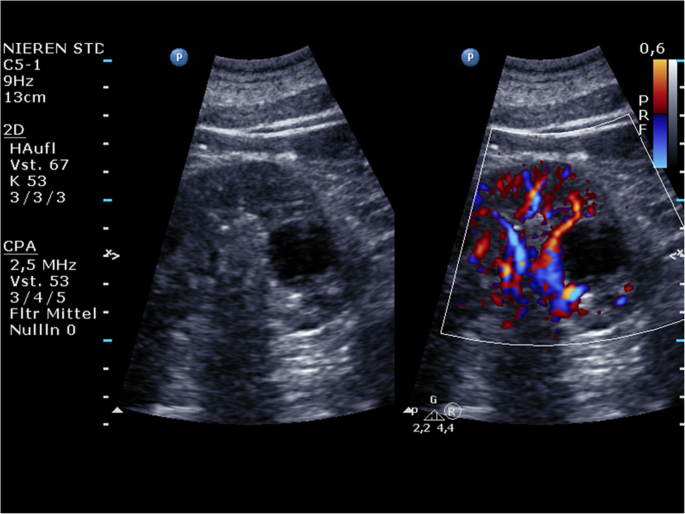
Philips Healthcare CX50: A renal cyst (left) that shows thin walls without any septa, calcifications, solid components, or contrast enhancement. There is no vascularisation of the cyst visible in colour Doppler mode (right)
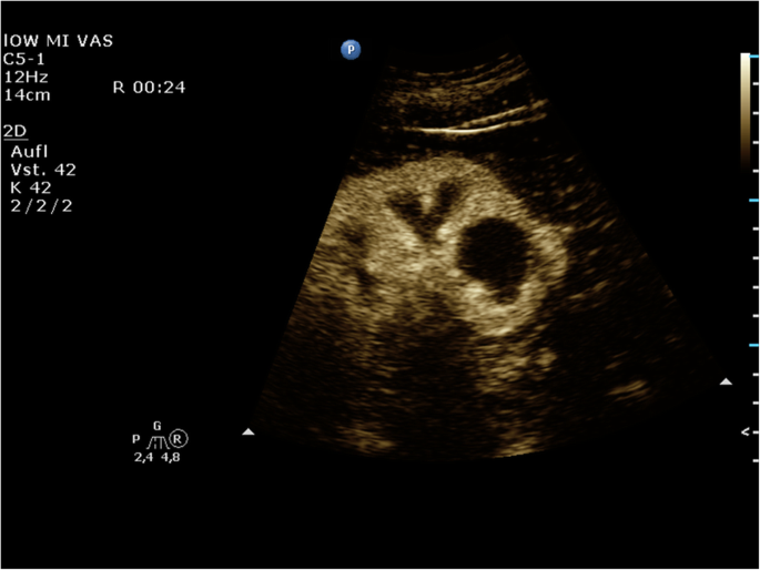
Philips Healthcare CX50: No contrast enhancement of the cyst can be detected; thus, the cyst can be classified as Bosniak type I
Handheld ultrasound devices
Handheld ultrasound devices (HHUSD) can be taken anywhere and perfectly fit into a physician’s coat pocket. Many companies produce HHUSDs. Examples include:
Fujifilm’s SonoSite iViz features a one-handed user interface that connects to a phased, curved, or linear transducer, while Doppler is also available. Stored studies can be exported via USB, DICOM, email, or uploaded to the cloud.
Clarius offers wireless HHUSDs that run with iOS and Android. Clarius Clip-Ons allow the user to have a multi-purpose scanner in one device.
The following sections describe the features of specific devices, as examples of succeeding generations of HHUSDs. They are not intended to imply that the described devices are the only, or the best example in each category, but have been chosen simply as illustrative examples.
The first generation of HHUSD: Acuson P10 Siemens
The Acuson P10 system by Siemens Medical Solutions was introduced in 2007 as the first portable ultrasound device. It weighs about 0.730 kg and was designed for immediate and easy use in emergency medicine, cardiology, and obstetrics with an intuitive Personal Digital Assistant (PDA) interface. It features a 3.7-inch touch-screen LCD display with 640 x 480 pixels with a wide viewing angle and a lithium battery which provides a run time of approximately 60 minutes. According to the manufacturer’s manual, the start-up time is 10 seconds. The Acuson P4-2 phased array transducer allows abdominal, renal, obstetric, transthoracic, and cardiac applications in the context of emergency medicine. The frequency range is 2–4 Mhz and allows for 2–24 cm of display depth with up to 28 frames per second. Examinations can be stored on an SD memory card up to 2 GB or transferred via USB. The Acuson P10 offers 2D-mode imaging in fundamental and harmonic modes (Fig. 3 ). Besides abdominal diagnostic use, P10 ultrasound devices were used in cardiac imaging [ 4 , 15 , 16 , 17 , 18 ].
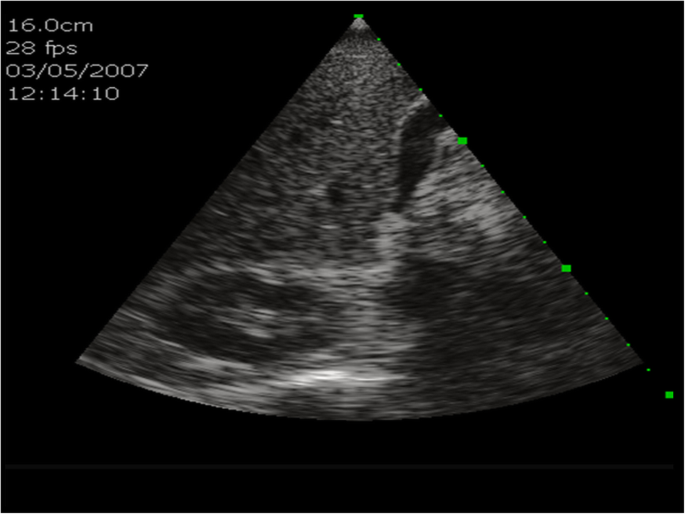
Acuson P10 Siemens: B-scan ultrasound of the liver, including the gall bladder and the right kidney
The second generation of HHUSD: GE Healthcare VScan with Dual Probe
The recent GE Healthcare VScan with Dual Probe is the first kind of ultrasound devices that features two transducers in a single probe. The VScan with Dual Probe is designed for a broad spectrum of clinical applications, including ultrasound-guided catheter placements, cardiac, abdominal, thoracic, and foetal issues. It weighs 0.436 kg and has a 3.5-inch display allowing for 240 x 320 pixels resolution. The phased array transducer allows for a 75-degree field with a maximum depth of 24 cm for B-mode and for colour Doppler mode with an angle of up to 40 degrees. The frequency range is 1.7–3.8 MHz. The linear array transducer allows for a maximum depth of 8 cm for B-mode and for colour Doppler mode. The frequency range for the linear array transducer is 3.4–8.0 MHz. Data can be stored on microSD (HC) cards or transferred via USB. According to the manufacturer’s manual, the battery runtime is about 60 minutes. Besides abdominal diagnostic use, Vscan ultrasound devices were used in MSK, cardiac imaging, OB/GYN, and emergency settings [ 13 , 19 , 20 , 21 , 22 , 23 , 24 , 25 ] (Figs. 4 , 5 ).
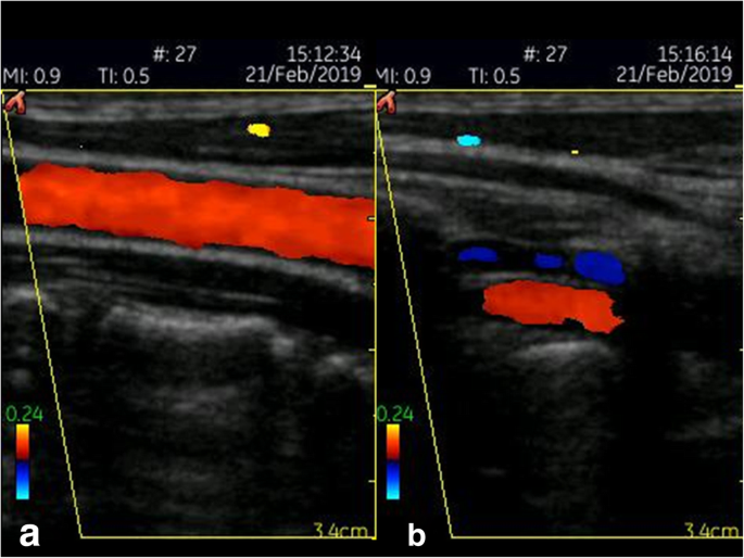
GE Healthcare VScan (linear array transducer): Colour Doppler image of the carotid ( a ) and vertebral artery ( b )
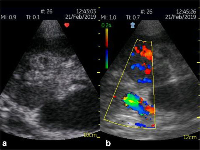
GE Healthcare VScan (phased array transducer): B-scan ultrasound ( a ) and colour Doppler ( b ) of a transplant kidney
The third generation of HHUSD: Philips Lumify
The Philips ultrasound Lumify works with a compatible smart device (e.g., smartphone or tablet). Using the Lumify, three different transducers are available: an S4-1 broadband sector array (4 to 1 MHz), a C5-2 broadband curved array transducer (5 to 2 MHz) and an L12-4 broadband linear array transducer (12 to 4 MHz). The system allows 2D, steerable colour Doppler, M-mode, advanced XRES, and multivariate harmonic imaging and SonoCT. By using different transducers, applications can be extended to include Cardiac, OB/GYN, Lung, Abdomen, FAST, Soft Tissue, Vascular, Superficial, and Musculoskeletal. For the first time, a compatible smartphone or mobile ultrasound device can be used to plug in an ultrasound transducer. All necessary beamforming and data management is done in the probe. The mobile smart device is only necessary for the battery supply and the display. The Lumify app must be uploaded on the mobile device to enhance imaging. Advanced imaging algorithms are automatically available and create the image. By using the touch screen of the mobile device, depth, gain, power, and colour can be optimised.
In a study by Miller et al, 56 patients scheduled for either reconstructive or aesthetic surgery were evaluated preoperatively and/or intraoperatively by a single surgeon with a linear 12–4 probe using the Philips Lumify device. For patients undergoing flap reconstruction, potential donor sites were imaged in order to locate the largest perforator. For patients undergoing abdominal procedures, intraoperative visualisation of the abdominal muscular layers was used for the delivery of anaesthesia during transversus abdominis plane block. Lastly, the superficial fascial system was subjectively evaluated in all preoperative patients. The conclusion of the study was that the newest, miniaturised colour Doppler ultrasound technology has a variety of applications that may improve patient outcomes and experience in plastic surgery [ 26 ]. Besides abdominal diagnostic use, the Lumify and the Visiq ultrasound devices were used in abdominal imaging, emergency, and general imaging [ 26 , 27 , 28 , 29 ] (Figs. 6 , 7 ).
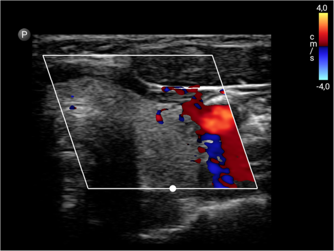
Philips Lumify linear transducer: Colour Doppler of the left thyroid lobe including the carotid artery
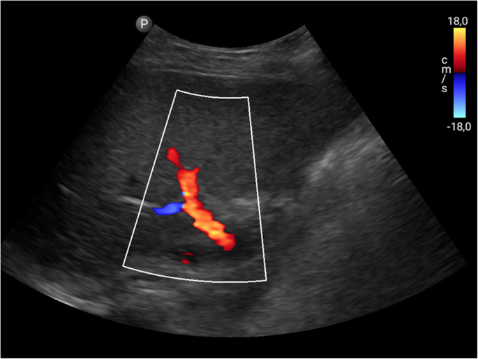
Philips Lumify curved array transducer: Colour Doppler imaging of the portal vein
Outlook: Butterfly’s iQ
As a very recent innovative start-up, the Butterfly Network's handheld ultrasound device features a silicon chip (2D array, 9000 micro-machined sensors) instead of piezoelectric crystals to induce ultrasound waves (“Ultrasound-on-Chip technology”). This allows it to emulate curved, linear, or phased transducers at any time in M-, B-mode or colour Doppler with 2–30 cm scan depth. It weighs only 0.313 kg and is connected to a smartphone. The battery run time is 120 minutes and the wireless full recharge takes up to 5 hours. Moreover, the ultrasound findings can be uploaded to the Butterfly Cloud, so any expert with access can help evaluate the sonographic findings. By using artificial intelligence algorithms, the position of the probe can be adjusted to meet the requirements of the user (Figs. 8 , 9 ).
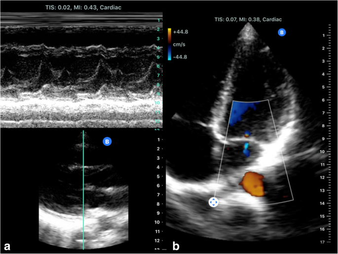
Butterfly’s iQ emulate phased transducers: Cardiac imaging including M-Mode ( a ) and four chamber view with colour Doppler ( b )
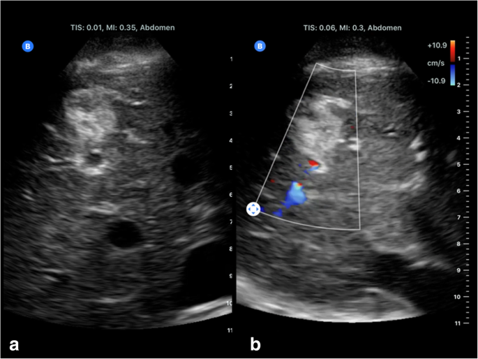
Butterfly’s iQ emulate curved transducers: B-scan ultrasound ( a ) and colour Doppler ( b ) of a liver haemangioma
Healthcare workers or paramedics might be equipped with handheld ultrasound devices like Butterfly’s iQ and artificial guidance with the immediate ultrasound correlates with rapidly recognising serious health issues. In the future, even patients might be provided with handheld ultrasound devices, so their caring physicians might, without directly seeing the patient, evaluate uploaded ultrasound findings. Furthermore, healthcare systems in developing countries may benefit immensely from affordable ultrasound devices [ 30 , 31 ].
Hand-held devices open up new possibilities of imaging at the point of care, in whichever setting this is needed. However, adequate training of ultrasound users, image and report storage for further reference, and high standards of hygiene are mandatory. Patient safety must not be compromised.
The newest, miniaturised handheld ultrasound devices technology has a variety of applications that may improve patient outcomes and experience [ 32 ]. The overall time required for performing an ultrasound examination at the bedside can be considerably reduced if a portable device is used instead of a mobile system [ 33 ]. High standards of hygiene must be maintained. Images and reports must be stored in patient records for further reference.
Availability of data and materials
Not applicable.
Busch M (2006) Portable ultrasound in pre-hospital emergencies: a feasibility study. Acta Anaesthesiol Scand 50(6):754–758
Article CAS Google Scholar
Price DD, Wilson SR, Murphy TG (2000) Trauma ultrasound feasibility during helicopter transport. Air Med J 19(4):144–146
Kobal SL, Trento L, Baharami S, et al (2005) Comparison of effectiveness of hand-carried ultrasound to bedside cardiovascular physical examination. Am J Cardiol 96(7):1002–1006
Article Google Scholar
Stock KF, Klein B, Steubl D, et al (2015) Comparison of a pocket-size ultrasound device with a premium ultrasound machine: diagnostic value and time required in bedside ultrasound examination. Abdom Imaging 40(7):2861–2866
Nyhsen CM, Humphreys H, Koerner RJ, et al (2017) Infection prevention and control in ultrasound - best practice recommendations from the European Society of Radiology Ultrasound Working Group. Insights Imaging 8(6):523–535
European Society of Radiology (ESR); European Federation of Societies for Ultrasound in Medicine and Biology (EFSUMB) (2013 Oct) Joint ESR-EFSUMB recommendation on archiving and reporting ultrasound examinations. Insights Imaging 4(5):525–526
Stawicki SP, Howard JM, Pryor JP, et al (2010) Portable ultrasonography in mass casualty incidents: The CA-VEAT examination. World J Orthop 1(1):10–19
Vourvouri EC, Poldermans D, Schinkel AF, et al (2001) Abdominal aortic aneurysm screening using a hand-held ultrasound device. “A pilot study”. Eur J Vasc Endovasc Surg 22(4):352–354
Zhou JY, Rappazzo KC, Volland L, et al (2016) Pocket handheld ultrasound as a novel point-of-care imaging modality to diagnose bleeding in hemophilic joints. Blood 128:2345
McGahan JP, Pozniak MA, Cronan J, et al (2015) Handheld ultrasound: Threat or opportunity? Appl Radiol 44(3):20-25
Winters KD, Toth-Manikowski S, Martire C, Shafi T (2016) Hand-carried ultrasound use in clinical nephrology: Case report. Medicine (Baltimore) 95(30):e4166
Keil-Ríos D, Terrazas-Solís H, González-Garay A, Sánchez-Ávila JF, García-Juárez I (2016) Pocket ultrasound device as a complement to physical examination for ascites evaluation and guided paracentesis. Intern Emerg Med 11(3):461–466
Michon A, Jammal S, Passeron A, et al (2019) Use of pocket-sized ultrasound in internal 381 medicine (hospitalist) practice: Feedback and perspectives. Rev Med Intern 40(4):220-225
Oschatz E, Prosch H, Schober E, Mostbeck G (2004) Evaluation of a portable ultrasound device immediately after spiral computed tomography. Ultraschall Med 25:433–437
Kimura BJ, Gilcrease GW, Showalter BK, Phan JN, Wolfson T (2012) Diagnostic performance of a pocket-sized ultrasound device for quick-look cardiac imaging. Am J Emerg Med 30(1):32–36
Dalla Pozza R, Loeff M, Kozlik-Feldmann R, Netz H (2010) Hand-carried ultrasound devices in pediatric cardiology: clinical experience with three different devices in 110 patients. J Am Soc Echocardiogr 23(12):1231–1237
Culp BC, Mock JD, Chiles CD, Culp WC Jr (2010) The pocket echocardiograph: validation and feasibility. Echocardiography 27(7):759–764
Egan M, Ionescu A (2008) The pocket echocardiograph: a useful new tool? Eur J Echocardiogr 9(6):721–725
PubMed Google Scholar
Lau BC, Motamedi D, Luke A (2018) Use of Pocket-Sized Ultrasound Device in the Diagnosis of Shoulder Pathology. Clin J Sport Med. https://doi.org/10.1097/JSM.0000000000000577
Sisó-Almirall A, Kostov B, Navarro González M, et al (2017) Abdominal aortic aneurysm screening program using hand-held ultrasound in primary healthcare. PLoS One 12(4):e0176877
Colclough A, Nihoyannopoulos P (2017) Pocket-sized point-of-care cardiac ultrasound devices Role in the emergency department. Herz 42(3):255–261
Johnson GG, Zeiler FA, Unger B, Hansen G, Karakitsos D, Gillman LM (2016) Estimating the accuracy of optic nerve sheath diameter measurement using a pocket-sized, handheld ultrasound on a simulation model. Crit Ultrasound J 8(1):18
Bruns RF, Menegatti CM, Martins WP, Araujo Júnior E (2015) Applicability of pocket ultrasound during the first trimester of pregnancy. Med Ultrason 17(3):284–288
Ruddox V, Stokke TM, Edvardsen T, et al (2013) The diagnostic accuracy of pocket-size cardiac ultrasound performed by unselected residents with minimal training. Int J Cardiovasc Imaging 29(8):1749–1757
Kitada R, Fukuda S, Watanabe H, et al (2013) Diagnostic accuracy and cost-effectiveness of a pocket-sized transthoracic echocardiographic imaging device. Clin Cardiol 36(10):603–610
Miller JP, Carney MJ, Lim S, Lindsey JT (2018) Ultrasound and Plastic Surgery: Clinical Applications of the Newest Technology. Ann Plast Surg 80(6S Suppl 6:S356–S361
Nolting L, Baker D, Hardy Z, Kushinka M, Brown HA (2019) Solar-Powered Point-of-Care Sonography: Our Himalayan Experience. J Ultrasound Med. https://doi.org/10.1002/jum.14923
Maetani TH, Schwartz C, Ward RJ, Nissman DB (2018) Enhancement of Musculoskeletal Radiology Resident Education with the Use of an Individual Smart Portable Ultrasound Device (iSPUD). Acad Radiol 25(12):1659–1666
Zimmermann H, Rübenthaler J, Rjosk-Dendorfer D, et al (2015) Comparison of portable ultrasound system and high end ultrasound system in detection of endoleaks. Clin Hemorheol Microcirc 63(2):99–111
Butterfly iQ, a Whole Body Ultrasound That Fits in a Pocket, (2017). https://www.medgadget.com/2017/10/butterfly-iq-whole-body-ultrasound-fits-pocket.html . Accessed 17 July 2019
New “Ultrasound on a Chip” Tool Could Revolutionize Medical Imaging (2017) https://spectrum.ieee.org/the-human-os/biomedical/imaging/new-ultrasound-on-a-chip-tool-could-revolutionize-medical-imaging . Accessed 17 July 2019
Nelson BP, Melnick ER, Li J (2011) Portable ultrasound for remote environments, part II: current indications. J Emerg Med 40(3):313–321
Fischer T, Filimonow S, Petersein J, et al (2002) Ultrasound at the bedside: Does a portable ultrasound device save time? Ultraschall Med 23:311–314
Download references
Acknowledgments
This paper was prepared on behalf of the ESR Ultrasound Subcommittee by Dirk-Andre Clevert (Chair of the ESR Ultrasound Subcommittee) and Vincent Schwarze (Interdisciplinary Ultrasound-Center, Department of Radiology, University of Munich-Grosshadern Campus, Munich, Germany) with contributions from Christiane Nyhsen, Mirko D’Onofrio and Paul Sidhu (members of the ESR Ultrasound Subcommittee), and Adrian P. Brady (Chair of the ESR Quality, Safety and Standards Committee). The ESR Patient Advisory Group (ESR-PAG) reviewed and endorsed the paper in May 2019, and contributed the Patient Summary paragraph.
It was approved by the ESR Executive Council on July 11, 2019.
Author information
Authors and affiliations.
- Dirk-Andre Clevert
- , Vincent Schwarze
- , Christiane Nyhsen
- , Mirko D’Onofrio
- , Paul Sidhu
- & Adrian P. Brady
Contributions
DAC: conception and design of the paper, manuscript drafting and review. VS: manuscript drafting and review. All contributors reviewed and approved the final manuscript.
Ethics declarations
Ethics approval and consent to participate, consent for publication, competing interests.
M.D. discloses speaker’s honoraria by Bracco and Siemens, research grant/other research support by Hitachi, congress participation with Bracco and Siemens, and position on the consultant/advisory board of Bracco, Siemens and Hitachi. D.A.C. discloses speaker’s honoraria by Bracco, Siemens, Samsung and Philips and position on the consultant/advisory board of Bracco, Siemens, Samsung and Philips.
All other authors declare no competing interests.
Additional information
Publisher’s note.
Springer Nature remains neutral with regard to jurisdictional claims in published maps and institutional affiliations.
Rights and permissions
Open Access This article is distributed under the terms of the Creative Commons Attribution 4.0 International License ( http://creativecommons.org/licenses/by/4.0/ ), which permits unrestricted use, distribution, and reproduction in any medium, provided you give appropriate credit to the original author(s) and the source, provide a link to the Creative Commons license, and indicate if changes were made.
Reprints and permissions
About this article
Cite this article.
European Society of Radiology (ESR). ESR statement on portable ultrasound devices. Insights Imaging 10 , 89 (2019). https://doi.org/10.1186/s13244-019-0775-x
Download citation
Received : 17 July 2019
Accepted : 23 July 2019
Published : 16 September 2019
DOI : https://doi.org/10.1186/s13244-019-0775-x
Share this article
Anyone you share the following link with will be able to read this content:
Sorry, a shareable link is not currently available for this article.
Provided by the Springer Nature SharedIt content-sharing initiative
- Portable ultrasound devices
- Hand-carried ultrasound
- Ultrasound diagnostics

IMAGES
VIDEO
COMMENTS
The recommended parameters are. based on the Omnisound ultrasound machine and because this machine tends to have the better. BNR of 1:8:1 and an ERA of 5.0 cm2.29 Based on a study by Johns et al.,31 Five ultrasound. machines were tested including Chattanooga, Dynatron, 2 Omnisounds and XLTEX.
Ensure that your essay is well-organized, structured, descriptive, and engaging. To write an opinion essay on the topic, start with a clear and concise thesis statement, use specific examples and experiences to support your argument, address potential counterarguments, and use clear language that is easy for readers to understand.
Most theses in will have five chapters; (1) Introduction: statement of the problem, (2) review of literature, (3) design of study or methodology, (4) analysis of results and (5) summary, conclusions, and recommendations. Please note that the titles of each chapter are as they should be in the actual thesis.
Use of Ultrasound-Guidance for Arterial Puncture. All the anthropometric and demographic variables were recorded, as well as the main diagnosis of admission, comorbidities, the placement of the central venous catheter, and the course of the procedure. We will write a custom essay specifically for you by our professional experts.
The Department of circulation and medical imaging offers projects and master's thesis topics for technology students of most of the different technical study programmes at NTNU. ... Ultrasound Mediated Drug Delivery Ultrasound has very interesting applications for increasing transport of drugs to cancer tumors and also transport of genes into ...
i. Modifying ultrasound waveform parameters to control, influence, or disrupt cells. Thesis by. David Reza Mittelstein. In Partial Fulfillment of the Requirements for the degree of Doctor of Philosophy. CALIFORNIA INSTITUTE OF TECHNOLOGY Pasadena, California 2020 (Defended February 26, 2020) ii.
The job outlook for ultrasound technicians is positive, with the Bureau of Labor Statistics projecting a 12% growth in employment from 2019 to 2029, much faster than the average for all occupations. This growth is driven by an aging population that will require more medical imaging services for diagnosis and treatment.
While ultrasound is useful for noninvasively locating suspicious lesions in organs such as the breast and prostate, a tissue biopsy is required to make an accurate diagnosis in most cases. The majority of biopsies are benign, and the procedures are typically invasive and uncomfortable (e.g., a typical prostate biospy involves the removal of 12 ...
Acute abdomen is a medical emergency with a wide spectrum of etiologies. Point-of-care ultrasound (POCUS) can help in early identification and management of the causes. The ACUTE-ABDOMEN protocol was created by the authors to aid in the evaluation of acute abdominal pain using a systematic sonographic approach, integrating the same core ultrasound techniques already in use—into one mnemonic.
If you are a student studying ultrasound technology or a healthcare professional looking to expand your knowledge, here are 106 ultrasound essay topic ideas and examples to help you explore this fascinating field further. The history and development of ultrasound technology. The physics behind ultrasound imaging.
This thesis aims to assess current ultrasound tools and protocols for the diagnosis and management of ovarian tumours. I hypothesized that the IOTA (International Ovarian Tumour Analysis) tools would have less diagnostic accuracy when performed by a less ... Declaration Statement of Originality and Personal Contribution to Work_____ 2
Ultrasound Is a Supersonic Transmitter. This also serves as an important catalytic effects for the bonding of mothers to their babies even before they are born. It is also called the "re-assurance scans" and misnamed as "entertainment scans." Some specialists do not recommend 3-D and 4-D ultrasound as manadatory development of the conventional ...
The first step in ultrasound-guided nerve block is to visualize all the anatomical structures in the target area. All adjustable ultrasound variables, i.e. penetration depth, the frequencies, and the position of the focal zones, must be optimized for the type of block to be performed. Both the skin and the ultrasound probe need to be disinfected.
In the first trimester, pelvic ultrasound is employed to establish the presence or absence of an intrauterine gestational sac and to evaluate the viability of the pregnancy. In addition, it can be used to evaluate ectopic pregnancy and other pregnancy-related complications. Practice parameters for the performance and recording of obstetric ...
Ultrasound Imaging - Current Topics. Edited by: Felix Okechukwu Erondu. ISBN 978-1-78984-877-9, eISBN 978-1-78985-186-1, PDF ISBN 978-1-78985-331-5, Published 2022-05-11 Ultrasound Imaging - Current Topics presents complex and current topics in ultrasound imaging in a simplified format. It is easy to read and exemplifies the range of ...
Diagnostic ultrasounds use sound waves to make pictures of the body. Ultrasound, also called sonography, shows the structures inside the body. The images can help guide diagnosis and treatment for many diseases and conditions. Most ultrasounds are done using a device outside the body. However, some involve placing a small device inside the body.
In North Carolina as of 2012, there was 1770 people employed as a Ultrasound tech. In United States as of 2012 , there were 58,800 people employed as a ultrasound technician. The projected number of people employed as a sonographer in 2022 will be 85,900, employment is projected to grow 14-19%.
Step 2: Write your initial answer. After some initial research, you can formulate a tentative answer to this question. At this stage it can be simple, and it should guide the research process and writing process. The internet has had more of a positive than a negative effect on education.
Ultrasound-based elastography has rapidly replaced the need for liver biopsy in most patients with chronic liver disease in recent years. The technique is now widely supported by many manufacturers. This review will introduce various current ultrasound-based elastography techniques, review the physics and scanning techniques, discuss potential cofounding factors as well as summarising the ...
Portable ultrasound devices can be subdivided into three groups: laptop-associated devices, hand-carried (HCU), and handheld (HHUSD) systems. Almost all companies we investigated offer at least one portable ultrasound device. The big advantages of PUD lie in time saving (booting time, transfer, bedside positioning), e.g., at the bedside or in ...
Example 1: In a biochemistry class, you've been asked to write an essay explaining the impact of bisphenol A on the human body. Your thesis statement might say, "This essay will make clear the correlation between bisphenol A exposure and hypertension.". Check Circle.